
Page 794 - Clinical Immunology_ Principles and Practice ( PDFDrive )
P. 794
CHaPtEr 56 Inflammatory Muscle Diseases 765
TABLE 56.6 Differential Diagnosis of
Idiopathic Inflammatory Myopathy
Neuromuscular Disorders
Genetic muscular dystrophies
Metabolic myopathies
Disorders of carbohydrate metabolism: McArdle disease,
phosphofructokinase deficiency, adult acid maltase deficiency, and
others
Disorders of lipid metabolism: carnitine deficiency, carnitine palmitoyl
transferase deficiency
Disorders of purine metabolism: myoadenylate deaminase deficiency
Mitochondrial myopathies
Spinal muscular atrophies
Neuropathies: Guillain–Barré and other autoimmune polyneuropathies,
diabetes mellitus, porphyria
Myasthenia gravis and Eaton–Lambert syndrome
Amyotrophic lateral sclerosis
Myotonic dystrophy and other myotonias
Familial periodic paralysis
Endocrine and Electrolyte Disorders
Hypokalemia, hypercalcemia, hypocalcemia, hypomagnesemia
Hypothyroidism, hyperthyroidism
Cushing syndrome, Addison disease
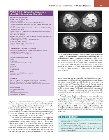
FIG 56.2 Magnetic Resonance Images of the Upper and Lower
toxic Myopathies (Partial List) Thighs of a Patient With Dermatomyositis Using the Fat-
Alcohol Suppressed T2 (STIR) Technique. With this technique inflam-
Amiodarone mation appears as a bright signal; normal muscle is gray; bone,
Chloroquine and hydroxychloroquine fat, fascia, and normal skin are dark. Blood vessels may appear
Cocaine as bright spots. Note the remarkable symmetry of the inflam-
Colchicine mation. In this patient most of the involvement is in the quadriceps
Corticosteroids in the upper thighs and around the periphery of the hamstring
D-penicillamine muscle group.
Ipecac
Statins and other lipid-lowering agents
Zidovudine (AZT)
37
Infections fascial tissue that was undetectable on clinical examination.
Viral: HIV, human T-lymphotropic virus 1 (HTLV-1), influenza MRI can also help differentiate active disease from chronic disease,
Bacterial: Staphylococcus, Streptococcus, Clostridia with active myositis being notable for changes consistent with
Parasitic: toxoplasmosis, trichinosis, schistosomiasis, cysticercosis muscle edema on T2-weighted images, and chronic myositis
revealing a decrease in muscle bulk and replacement by adipose
Miscellaneous on T1-weighted images. Although not specific, the changes of
38
Polymyalgia rheumatica inflammatory myopathy on imaging can provide considerable
Vasculitis
Eosinophilia myalgia syndrome assistance in confusing cases, as well as help in choosing a site
Paraneoplastic syndromes to biopsy.
A muscle biopsy should be performed in every suspected case
of myositis (Fig. 56.3). Although the patchy involvement means
that the biopsy can occasionally miss inflammation, certain
36
ESR, even with active disease. Likewise, hematological abnormali- confounding diagnoses—for example amyloidosis, eosinophilic
ties, including anemia, are uncommon and rarely related to the myositis, dystrophy, or some metabolic myopathies as well as
underlying myopathy. If a significant abnormality is found, the the important variant IBM—can be diagnosed definitively only
physician should be alert to another cause for it. by biopsy. The identification of autoantibodies, particularly the
Electromyographic (EMG) abnormalities are frequently myositis-specific autoantibodies, has distinct clinical and prog-
present. Although the test is useful for excluding some neurological nostic use.
diseases that resemble IIM, it is painful for many patients and
not useful for following the course of the illness.
MRI, especially a combination of the T1 and the fat-suppressed ON tHE HOrIZON
T2 (STIR) sequences, is remarkably useful in defining the extent • Development of revised and updated classification criteria for inflam-
of involvement and in planning a biopsy (Fig. 56.2). Whole-body matory myopathies is needed.
MRI has been shown to facilitate the characterization of inflam- • Improved diagnostic classification of cases through novel autoantibodies
matory myopathy, as certain patterns of muscle and subcutaneous as well as immunohistochemistry.
tissue inflammation were predictive of the IIM subset (DM, PM, • Long-term studies are needed to better characterize the prognosis of
or IBM). In a recent study in juvenile DM, whole-body MRI idiopathic inflammatory myopathies, particularly in relation to recently
discovered autoantibodies.
was able to reveal inflammatory changes of subcutaneous and