
Page 795 - Clinical Immunology_ Principles and Practice ( PDFDrive )
P. 795
766 Part SIX Systemic Immune Diseases
A B
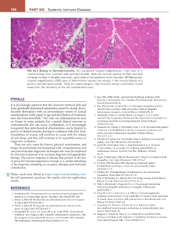
FIG 56.3 Biopsy in Dermatomyositis. (A) Low-power (original magnification ×100) view of a
muscle biopsy from a patient with dermatomyositis. Note the marked variation in fiber size and
the large number of atrophic myocytes, particularly at the periphery of the fascicles. (B) High-power
(original magnification ×200) view of inflammation around the vessels in the muscle biopsy of a
patient with dermatomyositis. There are nearby atrophic cells and cells whose nuclei have moved
away from the periphery of the cell (centralized nuclei).
5. Rose MR. 188th ENMC International Workshop: Inclusion Body
PITFALLS Myositis, 2–4 December 2011, Naarden, The Netherlands. Neuromuscul
Disord 2013;23:1044–55.
It is increasingly apparent that the boundary between IIM and 6. Kao AH, Lacomis D, Lucas M, et al. Anti-signal recognition particle
some genetically determined myopathies cannot be cleanly drawn. autoantibody in patients with and patients without idiopathic
Recently dystrophies with an extraordinary variety of clinical inflammatory myopathy. Arthritis Rheum 2004;50:209–15.
manifestations (with regard to age and distribution of weakness) 7. Christopher-Stine L, Casciola-Rosen LA, Hong G, et al. A novel
39
have also been described. Not only can inflammation be seen autoantibody recognizing 200-kd and 100-kd proteins is associated with
on biopsy in some patients, but a partial clinical response to an immune-mediated necrotizing myopathy. Arthritis Rheum
corticosteroids also can occur. Furthermore, it is increasingly 2010;62:2757–66.
recognized that mitochondrial abnormalities can be limited to 8. Mammen AL, Chung T, Christopher-Stine L, et al. Autoantibodies against
groups of skeletal muscles, leading to confusion with IIM. Toxic 3-hydroxy-3-methylglutaryl-coenzyme A reductase in patients with
myopathies, of course, will continue to occur with the release statin-associated autoimmune myopathy. Arthritis Rheum
2011;63:713–21.
of new drugs and thus will continue to be a possible source of 9. Lahouti AH, Amato AA, Christopher-Stine L. Inclusion body myositis:
diagnostic confusion. update. Curr Opin Rheumatol 2014;26:690–6.
Thus not only must the history, physical examination, and 10. Lloyd TE, Christopher-Stine L, Pinal-Fernandez I, et al. Cytosolic
biopsy be performed and interpreted with compulsiveness and 5’-nucleotidase 1A as a target of circulating autoantibodies in
care, but molecular diagnostic techniques also must be employed autoimmune diseases. Arthritis Care Res (Hoboken) 2016;68:
by clinicians in pursuit of an accurate diagnosis and appropriate 66–71.
therapy. The correct response to disease that persists in the face 11. Argov Z, Eisenberg I, Mitrani-Rosenbaum S. Genetics of inclusion body
of powerful immunosuppressive therapy is a careful rethinking myopathies. Curr Opin Rheumatol 1998;10:543–7.
of the diagnosis, including, on occasion, rebiopsy and molecular 12. Mathews MB, Bernstein RM. Myositis autoantibody inhibits
consultation. histidyl-tRNA synthetase: a model for autoimmunity. Nature
1983;304:177–9.
13. Dalakas MC. Pathophysiology of inflammatory and autoimmune
Please check your eBook at https://expertconsult.inkling.com/ myopathies. Presse Med 2011;40:e237–47.
for self-assessment questions. See inside cover for registration 14. Mor A, Wortmann RL, Mitnick HJ, et al. Drugs causing muscle disease.
details. Rheum Dis Clin North Am 2011;37:219–31, vi.
15. Walji S, Rubenstein J, Shannon P, et al. Disseminated pyomyositis
REFERENCES mimicking idiopathic inflammatory myopathy. J Rheumatol
2005;32:184–7.
1. Sontheimer RD. Dermatomyositis: an overview of recent progress with 16. Zong M, Loell I, Lindroos E, et al. Effects of immunosuppressive
emphasis on dermatologic aspects. Dermatol Clin 2002;20:387–408. treatment on interleukin-15 and interleukin-15 receptor alpha expression
2. Bohan A, Peter JB. Polymyositis and dermatomyositis (first of two parts). in muscle tissue of patients with polymyositis or dermatomyositis. Ann
N Engl J Med 1975;292:344–7. Rheum Dis 2012;71:1055–63.
3. Bohan A, Peter JB. Polymyositis and dermatomyositis (second of two 17. Greenberg SA, Pinkus JL, Pinkus GS, et al. Interferon-alpha/
parts). N Engl J Med 1975;292:403–7. beta-mediated innate immune mechanisms in dermatomyositis. Ann
4. Hoogendijk JE, Amato AA, Lecky BR, et al. 119th ENMC international Neurol 2005;57:664–78.
workshop: trial design in adult idiopathic inflammatory myopathies, with 18. Ikegame S, Takeda M, Ohno S, et al. Both RIG-I and MDA5 RNA
the exception of inclusion body myositis, 10–12 October 2003, Naarden, helicases contribute to the induction of alpha/beta interferon in measles
The Netherlands. Neuromuscul Disord 2004;14:337–45. virus-infected human cells. J Virol 2010;84:372–9.