
Page 789 - Clinical Immunology_ Principles and Practice ( PDFDrive )
P. 789
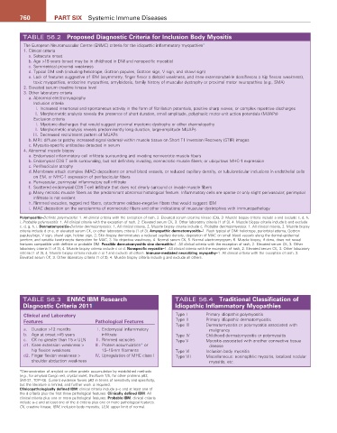
760          Part SIX  Systemic Immune Diseases


         TABLE 56.2  Proposed Diagnostic Criteria for Inclusion Body Myositis
          The European Neuromuscular Centre (ENMC) criteria for the idiopathic inflammatory myopathies 4
          1. Clinical criteria
           a. Subacute onset
           b. Age >18 years (onset may be in childhood in DM and nonspecific myositis)
           c. Symmetrical proximal weakness
           d. Typical DM rash (including heliotrope, Gottron papules, Gottron sign, V sign, and shawl sign)
           e. Lack of features suggestive of IBM (asymmetry, finger flexor ≥ deltoid weakness, and knee extensors/ankle dorsiflexors ≥ hip flexors weakness),
             toxic myopathies, endocrine myopathies, amyloidosis, family history of muscular dystrophy or proximal motor neuropathies (e.g., SMA)
          2. Elevated serum creatine kinase level
          3. Other laboratory criteria
            a. Abnormal electromyography
             Inclusion criteria
               I.  Increased insertional and spontaneous activity in the form of fibrillation potentials, positive sharp waves, or complex repetitive discharges
              II.  Morphometric analysis reveals the presence of short duration, small amplitude, polyphasic motor-unit action potentials (MUAPs)
             Exclusion criteria
               I.  Myotonic discharges that would suggest proximal myotonic dystrophy or other channelopathy
              II.  Morphometric analysis reveals predominantly long duration, large-amplitude MUAPs
              III.  Decreased recruitment pattern of MUAPs
            b. MRI: diffuse or patchy increased signal (edema) within muscle tissue on Short T1 Inversion Recovery (STIR) images
            c. Myositis-specific antibodies detected in serum
          4. Abnormal muscle biopsy
            a. Endomysial inflammatory cell infiltrate surrounding and invading nonnecrotic muscle fibers
            b. Endomysial CD8 T cells surrounding, but not definitely invading, nonnecrotic muscle fibers; or ubiquitous MHC-1 expression
            c. Perifascicular atrophy
            d. Membrane attack complex (MAC) depositions on small blood vessels, or reduced capillary density, or tubuloreticular inclusions in endothelial cells
             on EM, or MHC-1 expression of perifascicular fibers
            e. Perivascular, perimysial inflammatory cell infiltrate
            f.  Scattered endomysial CD8 T-cell infiltrate that does not clearly surround or invade muscle fibers
            g. Many necrotic muscle fibers as the predominant abnormal histological feature. Inflammatory cells are sparse or only slight perivascular; perimysial
             infiltrate is not evident
            h. Rimmed vacuoles, ragged red fibers, cytochrome oxidase–negative fibers that would suggest IBM
            i.  MAC deposition on the sarcolemma of nonnecrotic fibers and other indications of muscular dystrophies with immunopathology

        Polymyositis–Definite polymyositis: 1. All clinical criteria with the exception of rash, 2. Elevated serum creatine kinase (CK), 3. Muscle biopsy criteria include a and exclude c, d, h,
        i; Probable polymyositis: 1. All clinical criteria with the exception of rash, 2. Elevated serum CK, 3. Other laboratory criteria (1 of 3), 4. Muscle biopsy criteria include b and exclude
        c, d, g, h, i. Dermatomyositis–Definite dermatomyositis: 1. All clinical criteria, 2. Muscle biopsy criteria include c; Probable dermatomyositis: 1. All clinical criteria, 2. Muscle biopsy
        criteria include d or e, or elevated serum CK, or other laboratory criteria (1 of 3). amyopathic dermatomyositis–1. Rash typical of DM: heliotrope, periorbital edema, Gottron
        papules/sign, V sign, shawl sign, holster sign, 2. Skin biopsy demonstrates a reduced capillary density, deposition of MAC on small blood vessels along the dermal-epidermal
        junction, and variable keratinocyte decoration for MAC, 3. No objective weakness, 4. Normal serum CK, 5. Normal electromyogram, 6. Muscle biopsy, if done, does not reveal
        features compatible with definite or probable DM. Possible dermatomyositis sine dermatitis–1. All clinical criteria with the exception of rash, 2. Elevated serum CK, 3. Other
        laboratory criteria (1 of 3), 4. Muscle biopsy criteria include c or d. Nonspecific myositis–1. All clinical criteria with the exception of rash, 2. Elevated serum CK, 3. Other laboratory
        criteria (1 of 3), 4. Muscle biopsy criteria include e or f and exclude all others. Immune-mediated necrotizing myopathy–1. All clinical criteria with the exception of rash, 2.
        Elevated serum CK, 3. Other laboratory criteria (1 of 3), 4. Muscle biopsy criteria include g and exclude all others.


         TABLE 56.3  ENMC IBM research                           TABLE 56.4  traditional Classification of
         Diagnostic Criteria 2011  5                             Idiopathic Inflammatory Myopathies

          Clinical and Laboratory                                Type I     Primary idiopathic polymyositis
          Features                  Pathological Features        Type II    Primary idiopathic dermatomyositis
                                                                 Type III   Dermatomyositis or polymyositis associated with
          a.  Duration >12 months     I.  Endomysial inflammatory            malignancy
          b.  Age at onset >45 years  infiltrate                 Type IV    Childhood dermatomyositis or polymyositis
          c.  CK no greater than 15 × ULN   II.  Rimmed vacuoles  Type V    Myositis associated with another connective tissue
          d1.  Knee extension weakness >    III.  Protein accumulation* or   disease
            hip flexion weakness      15–18-nm filaments         Type VI    Inclusion body myositis
          d2.  Finger flexion weakness >     IV. Upregulation of MHC class I  Type VII  Miscellaneous: eosinophilic myositis, localized nodular
            shoulder abduction weakness                                      myositis, etc.
        *Demonstration of amyloid or other protein accumulation by established methods
        (e.g., for amyloid Congo red, crystal violet, thioflavin T/S, for other proteins p62,
        SMI-31, TDP-43). Current evidence favors p62 in terms of sensitivity and specificity,
        but the literature is limited, and further work is required.
        Clinicopathologically defined IBM: clinical criteria include a–c and at least one of
        the d criteria plus the first three pathological features. Clinically defined IBM: All
        clinical criteria plus one or more pathological features. Probable IBM: clinical criteria
        include a–c and at least one of the d criteria plus one or more pathological features.
        CK, creatine kinase; IBM, inclusion body myositis; ULN, upper limit of normal.