
Page 874 - Clinical Immunology_ Principles and Practice ( PDFDrive )
P. 874
846 Part Seven Organ-Specific Inflammatory Disease
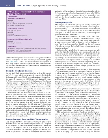
TABLE 62.1 Classification of Immune antibodies will be produced and can lead to significant hemolysis.
Hemolytic Disorders If the transplantation involves hematopoietic stem cells (HSCs),
this dilemma will resolve once the donor’s erythropoiesis pre-
autoimmune vails and the donor lymphocytes are no longer exposed to the
Warm Antibody-Mediated recipient RBCs.
Idiopathic
Secondary Immunopathogenesis
Drugs, lymphoid malignancies, infections
Other autoimmune diseases The antigens for antierythrocyte IgG are usually proteins, the
most clinically important of which are the Rh-associated glyco-
8
Cold Antibody-Mediated proteins (RhAG), D, C, c, E, and e. In contrast, antierythrocyte
Cold agglutinin disease IgM is directed at polysaccharides, which include the ABO and
Idiopathic I-antigens (I, i) found on the anion and glucose transporter
Secondary proteins in the RBC membrane. 9,10
Infection, lymphoid malignancies
Antibodies are distinguished by being “warm” and “cold”
Paroxysmal Cold Hemoglobinuria reactive, respectively, meaning that they bind antigens at core
Idiopathic body temperature (warm) or they bind antigens preferentially
Secondary to infections at lower temperatures (cold) in the peripheral circulation or ex
vivo. This distinction results from the different thermodynamics
alloimmune of binding to protein (hydrophobic) and polysaccharide (elec-
Secondary to red cell transfusions (alloantibodies, isoantibodies) trostatic) antigens. 10
Secondary to fetal-maternal hemorrhage IgG antibodies typically bind at warm temperatures and IgM
Secondary to transplanted lymphocytes antibodies typically bind at cold temperatures, although there
can be overlap and exceptions (e.g., the Donath-Landsteiner IgG
antibodies that are seen in paroxysmal cold hemoglobinuria).
children following a viral illness and can be managed by avoidance IgG and IgM also differ in their ability to fix complement, and
of cold. In the past, it was more commonly associated with syphilis this affects the resulting mechanism of hemolysis. To attach the
(see Table 62.1). There is also an autoimmune variety of PCH first component of the classical complement pathway, two IgG
that may require immunosuppression with corticosteroids. molecules must bind in close proximity on the RBC. However,
Splenectomy is not helpful as a consequence of the fact that the because of its pentameric structure, a single IgM molecule can
hemolysis is intravascular. initiate complement activation.
Erythrocyte-bound IgG becomes attached to the Fc receptors
Hemolytic Transfusion Reactions of splenic macrophages, which may engulf all or part of the cell
Because individuals with group O RBCs have preformed iso-anti-A or release lysosomal enzymes that digest its membrane (antibody-
11
and -B, they must only be transfused with group O cells. Similarly, dependent cell-mediated cytotoxicity [ADCC]). RBC fragments
individuals with group A RBCs with preformed anti-B isoantibod- escaping from this encounter lose more membrane than cytoplasm
ies must only receive group A RBCs, and individuals with group and become spherical (spherocytes) as a consequence of this
B RBCs must only receive group B RBCs. Because of the absence change in the surface-to-volume ratio. If IgG has initiated
of either group A or B antigens on the surface of group O RBCs, complement activation on the cell surface, binding of C3b to
such cells can be used in transfusion of A, B, or AB individuals splenic macrophages will augment erythrocyte phagocytosis in
in emergency situations. Failure to abide by these rules results the spleen. 12
in acute intravascular hemolysis that can cause renal failure, When IgM fixes complement, the process begins in the cooler
disseminated intravascular coagulation, and death. peripheral circulation, where IgM binds to RBCs. If the amount of
Other hemolytic transfusion reactions are caused by alloan- IgM bound is relatively high with at least some of it remaining on
tibodies, predominantly IgG. Therefore a multiply transfused the cell at 37°C (e.g., anti-A or anti-B isoantibodies), the cascade
patient is at risk for hemolysis from alloantibodies if the patient of complement activation goes to completion. Doughnut-shaped
receives incompatible blood. Multiparous women are at similar holes are formed in the cell membrane that allow the influx of
risk because of exposure to paternal fetal RBC antigens. Fortu- water and sodium, inducing intravascular osmotic rupture of
11
nately, for unclear reasons, most RBC antigens do not elicit an the cell. However, if the IgM elutes from the RBC as it returns
immune response although the Rh, Kell, Kidd, and Duffy antigens to body core temperature, the complement reaction attenuates.
are clearly immunogenic. Exposure particularly to Kidd or Duffy In this circumstance, the components remain on the cell but do
antigens may stimulate alloantibody formation that can rise to not cause intravascular hemolysis. Instead, the cells are cleared
sufficient titer to cause hemolysis with an onset typically delayed by hepatic macrophages via complement-binding sites. 13
by approximately 1 week. Such delayed transfusion reactions This has important implications for clinical tests for the pres-
may be subtle or cause an abrupt drop in hemoglobin with ence of antibody and complement on the RBC surface during
jaundice and hemoglobinuria. the workup of immune-mediated hemolysis. Antibody-mediated
hemolysis causes variable degrees of anemia and reticulocytosis.
Immune Hemolysis Associated With Transplantation Intravascular hemolysis releases hemoglobin into the circulation,
Any transplanted tissue may contain “passenger” lymphocytes but this is too small an amount to cause measurable hemoglo-
from the donor that will survive and proliferate in the recipient binemia, although it will result in consumption of haptoglobin,
7
if the recipient is sufficiently immunosuppressed. When the which is rapidly depleted. In contrast, when hemolysis results from
RBCs of the recipient carry A or B antigen and the donor is complement-mediated lysis, such as follows an ABO-incompatible
ABO incompatible, the transplanted lymphocytes will respond blood transfusion, hemoglobinemia becomes massive, overcom-
to the recipient RBCs as foreign, and allogeneic anti-A or anti-B ing the scavenging capacity of plasma hemoglobin binders