
Page 877 - Clinical Immunology_ Principles and Practice ( PDFDrive )
P. 877
CHaPter 62 Immunohematological Disorders 849
have been attempts in the past to classify this pattern as
pseudo-Felty syndrome, but the clinical findings and course
are often indistinguishable from “classic” Felty syndrome.
2. About half the patients with clinically apparent T-LGL leukemia
have circulating rheumatoid factor and immune complexes
in their blood; about a third, usually patients expressing
HLA-DR4, develop clinically significant arthritis, sometimes
requiring antiinflammatory agents.
Although the reason why clonal T-LGL disorders and autoim-
munity often coexist is unclear, the tendency toward overlap is
clear. Since the pathophysiology and therapy of both conditions
are similar, these problems in classification usually have little
impact on the initial management of neutropenia. When a patient
with “Felty syndrome” develops aggressive T-LGL leukemia or
a patient with T-LGL manifests severe rheumatological symptoms,
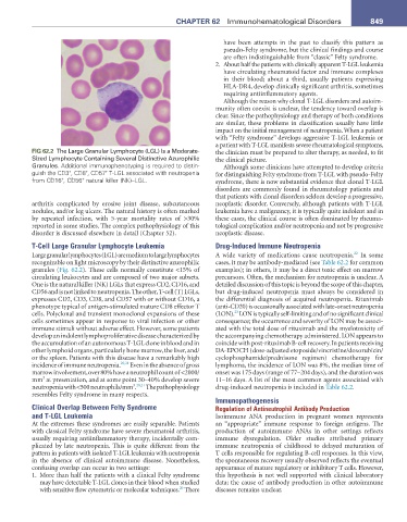
FIG 62.2 The Large Granular Lymphocyte (LGL) Is a Moderate- the clinician must be prepared to alter therapy, as needed, to fit
Sized Lymphocyte Containing Several Distinctive Azurophilic the clinical picture.
Granules. Additional immunophenotyping is required to distin- Although some clinicians have attempted to develop criteria
guish the CD3 , CD8 , CD57 T-LGL associated with neutropenia for distinguishing Felty syndrome from T-LGL with pseudo-Felty
+
+
+
from CD16 , CD56 natural killer (NK)–LGL. syndrome, there is now substantial evidence that clonal T-LGL
+
+
disorders are commonly found in rheumatology patients and
that patients with clonal disorders seldom develop a progressive,
arthritis complicated by erosive joint disease, subcutaneous neoplastic disorder. Conversely, although patients with T-LGL
nodules, and/or leg ulcers. The natural history is often marked leukemia have a malignancy, it is typically quite indolent and in
by repeated infection, with 5-year mortality rates of >30% these cases, the clinical course is often dominated by rheuma-
reported in some studies. The complex pathophysiology of this tological complication and/or neutropenia and not by progressive
disorder is discussed elsewhere in detail (Chapter 52). neoplastic disease.
T-Cell Large Granular Lymphocyte Leukemia Drug-Induced Immune Neutropenia
22
Large granular lymphocytes (LGL) are medium to large lymphocytes A wide variety of medications cause neutropenia. In some
recognizable on light microscopy by their distinctive azurophilic cases, it may be antibody-mediated (see Table 62.2 for common
granules (Fig. 62.2). These cells normally constitute <15% of examples); in others, it may be a direct toxic effect on marrow
circulating leukocytes and are composed of two major subsets. precursors. Often, the mechanism for neutropenia is unclear. A
One is the natural killer (NK) LGLs that express CD2, CD16, and detailed discussion of this topic is beyond the scope of this chapter,
CD56 and is not linked to neutropenia. The other, T-cell (T) LGLs, but drug-induced neutropenia must always be considered in
expresses CD2, CD3, CD8, and CD57 with or without CD16, a the differential diagnosis of acquired neutropenia. Rituximab
phenotype typical of antigen-stimulated mature CD8 effector T (anti-CD20) is occasionally associated with late-onset neutropenia
23
cells. Polyclonal and transient monoclonal expansions of these (LON). LON is typically self-limiting and of no significant clinical
cells sometimes appear in response to viral infection or other consequence; the occurrence and severity of LON may be associ-
immune stimuli without adverse effect. However, some patients ated with the total dose of rituximab and the myelotoxicity of
develop an indolent lymphoproliferative disease characterized by the accompanying chemotherapy administered. LON appears to
the accumulation of an autonomous T-LGL clone in blood and in coincide with post-rituximab B-cell recovery. In patients receiving
other lymphoid organs, particularly bone marrow, the liver, and/ DA-EPOCH (dose-adjusted etoposide/vincristine/doxorubicin/
or the spleen. Patients with this disease have a remarkably high cyclophosphamide/prednisone regimen) chemotherapy for
incidence of immune neutropenia. 20,21 Even in the absence of gross lymphoma, the incidence of LON was 8%, the median time of
marrow involvement, over 80% have a neutrophil count of <2000/ onset was 175 days (range of 77–204 days), and the duration was
3
mm at presentation, and at some point 30–40% develop severe 11–16 days. A list of the most common agents associated with
3 20,21
neutropenia with <500 neutrophils/mm . The pathophysiology drug-induced neutropenia is included in Table 62.2.
resembles Felty syndrome in many respects.
Immunopathogenesis
Clinical Overlap Between Felty Syndrome Regulation of Antineutrophil Antibody Production
and T-LGL Leukemia Isoimmune ANA production in pregnant women represents
At the extremes these syndromes are easily separable. Patients an “appropriate” immune response to foreign antigens. The
with classical Felty syndrome have severe rheumatoid arthritis, production of autoimmune ANAs in other settings reflects
usually requiring antiinflammatory therapy, incidentally com- immune dysregulation. Older studies attributed primary
plicated by late neutropenia. This is quite different from the immune neutropenia of childhood to delayed maturation of
pattern in patients with isolated T-LGL leukemia with neutropenia T cells responsible for regulating B-cell responses. In this view,
in the absence of clinical autoimmune disease. Nonetheless, the spontaneous recovery usually observed reflects the eventual
confusing overlap can occur in two settings: appearance of mature regulatory or inhibitory T cells. However,
1. More than half the patients with a clinical Felty syndrome this hypothesis is not well supported with clinical laboratory
may have detectable T-LGL clones in their blood when studied data; the cause of antibody production in other autoimmune
20
with sensitive flow cytometric or molecular techniques. There diseases remains unclear.