
Page 903 - Clinical Immunology_ Principles and Practice ( PDFDrive )
P. 903
CHaPtEr 64 Immunology of Psoriasis 873
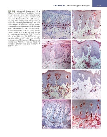
FIG 64.2 Histological Components of a
Mature Psoriatic Plaque. Psoriatic skin lesions
are characterized by a hyperproliferative epi-
dermis showing an increased mitotic rate of
the basal keratinocytes (A, Ki67 immuno-
staining). As a consequence, the epidermis is
thickened, with elongated rete ridges that form
typical finger-like protrusions into the dermis.
The epidermis becomes infiltrated by activated
CD8 T lymphocytes and neutrophils (B and C,
immunostaining for CD8 and CD15, respec-
tively). Within the drmis, an inflammatory
+
infiltrate mainly composed of CD3 T cells (D),
+
+
CD11c dendritic cells (E), BDCA-2 plasma-
+
cytoid dendritic cells (F), c-kit mast cells (G), A B
and neutrophils (C) is observed. Elongated and
+
dilated ICAM-1 (H) blood vessels in the dermis
represent another histological hallmark of
psoriatic skin.
C D
E F
G H