
Page 928 - Clinical Immunology_ Principles and Practice ( PDFDrive )
P. 928
896 Part Seven Organ-Specific Inflammatory Disease
of cortical lesions have been described: leukocortical (which span
gray and white matter), intracortical, and subpial. All of these
lesions show demyelination and oligodendrocyte loss, microglial
activation, neuritic transections, neuronal death, and reduced
presynaptic terminals. Subpial lesions are the most common.
They can cover long distances of the cortical ribbon and usually
extend to the cortical layer III or IV. Cortical lesions are not
visible on conventional MRI scans and require special staining
to be appreciated in CNS tissue sections. This explains why they
were not recognized as common features of MS until recently.
In fact, cortical demyelination and gray matter atrophy are evident
from the earliest stages of disease, even before a clinically definite
diagnosis can be made, and continue to advance at an increasing
rate throughout the disease course. Extensive cortical demyelin-
ation is evident in the forebrain and cerebellum during progressive
MS. Gray matter atrophy in individuals with MS correlates
strongly with cognitive deficits and clinical disability. 36
Meningeal Inflammation
White blood cell (WBC) counts tend to be within normal limits
or only slightly elevated in the CSF of most patients with MS.
Nonetheless, there is growing recognition that low-grade diffuse
meningeal inflammation and focal perivascular meningeal
inflammation are common. Meningeal inflammation is most
prominent in progressive forms of MS but is prevalent in early
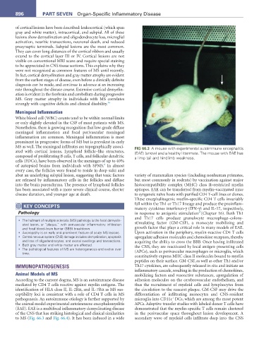
MS as well. The meningeal infiltrates are topographically associ- FIG 66.3 A mouse with experimental autoimmune encephalitis
ated with cortical lesions. Lymphoid follicle–like structures, (EAE) (arrow) and a healthy littermate. The mouse with EAE has
composed of proliferating B cells, T cells, and follicular dendritic a limp tail and hindlimb weakness.
cells (FDCs), have been observed in the meninges of up to 40%
6
of autopsied brains from individuals with SPMS. In almost
every case, the follicles were found to reside in deep sulci and
abut an underlying subpial lesion, suggesting that toxic factors variety of mammalian species (including nonhuman primates,
are released by inflammatory cells in the follicles and diffuse but most commonly in rodents) by vaccination against major
into the brain parenchyma. The presence of lymphoid follicles histocompatibility complex (MHC) class II–restricted myelin
has been associated with a more severe clinical course, shorter epitopes. EAE can be transferred from myelin-vaccinated mice
disease duration, and younger age at death. to syngeneic naïve hosts with purified CD4 T-cell lines or clones.
These encephalitogenic myelin-specific CD4 T cells invariably
KeY COnCePtS fall within the Th1 or Th17 lineage and produce the proinflam-
matory cytokines interferon-γ (IFN-γ) and IL-17, respectively,
Pathology in response to antigenic stimulation (Chapter 16). Both Th1
37
and Th17 cells produce granulocyte macrophage–colony-
• The hallmark of multiple sclerosis (MS) pathology is the focal demyelin-
ated lesion, or “plaque,” with perivascular inflammatory infiltration stimulating factor (GM-CSF), a monocyte mobilizing and
and focal blood–brain barrier (BBB) breakdown. growth factor that plays a critical role in many models of EAE.
• Axonopathy is an early and prominent feature of acute MS lesions. Upon activation in the periphery, myelin-reactive CD4 T cells
• Central nervous system (CNS) damage includes demyelination, apoptosis upregulate adhesion molecules and chemokine receptors, thereby
and loss of oligodendrocytes, and axonal swellings and transections. acquiring the ability to cross the BBB. Once having infiltrated
• Both gray matter and white matter are affected. the CNS, they are reactivated by local antigen-presenting cells
• The pathological features of MS are heterogeneous and evolve over
time. (APCs), such as perivascular macrophages or microglia, which
constitutively express MHC class II molecules bound to myelin
peptides on their surface. GM-CSF, as well as other Th1 and/or
IMMUNOPATHOGENESIS Th17 cytokines, are subsequently released in situ and initiate an
inflammatory cascade, resulting in the production of chemokines,
Animal Models of MS mobilizing factors and vasoactive substances, upregulation of
According to the current dogma, MS is an autoimmune disease adhesion molecules on the cerebrovascular endothelium, and
mediated by CD4 T cells reactive against myelin antigens. The thus the recruitment of myeloid cells and lymphocytes from
identification of HLA class II, IL-2Rα, and IL-7Rα as MS sus- the circulation to the nascent plaque. GM-CSF may drive the
ceptibility loci is consistent with a role of CD4 T cells in MS differentiation of infiltrating monocytes and CNS-resident
+
pathogenesis. An autoimmune etiology is further supported by microglia into CD11c DCs, which are among the most potent
the animal model experimental autoimmune encephalomyelitis APCs. Adoptive transfer studies with labeled donor T cells have
(EAE). EAE is a multifocal inflammatory demyelinating disease demonstrated that the myelin-specific T cells remain clustered
of the CNS that has striking histological and clinical similarities in the perivascular space throughout lesion development. A
to MS (Fig. 66.3 and Fig. 66.4). It has been induced in a wide secondary wave of myeloid cells infiltrate deep into the CNS