
Page 927 - Clinical Immunology_ Principles and Practice ( PDFDrive )
P. 927
CHaPter 66 Multiple Sclerosis 895
Sex Hormones
MS relapse rates decline during pregnancy, particularly in the
third trimester, and rebound in the first 3 months post partum
before returning to the prepregnancy rate. This has led to the
hypothesis that certain female sex hormones may play a protective
role in RRMS. Estriol is an estrogen unique to pregnancy. It is
synthesized by the fetoplacental unit and reaches its highest levels
in the last trimester. A randomized, double-blinded, placebo-
controlled phase II trial of estriol in combination with glatiramer
acetate (GA) versus placebo plus GA showed a reduction in the
32
annualized relapse rate at 2 years in the estriol-treated group.
Animal model studies have indicated that estrogens, including
estriol, have antiinflammatory and neuroprotective effects through
engagement of the estrogen receptors expressed on leukocytes
and CNS resident cells, respectively.
Testosterone has neuroprotective effects in animal models of
MS, and decreased testosterone levels in males with MS were
33
reported to be associated with disability. In a small, open-label,
phase II clinical trial, testosterone treatment appeared to arrest
loss of gray matter (and even to reverse atrophy of gray matter
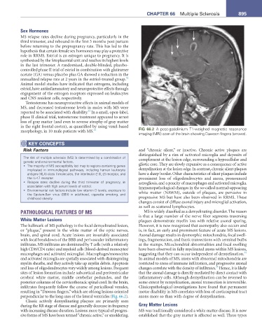
in the right frontal cortex), as quantified by using voxel-based FIG 66.2 A post-gadolinium T1-weighed magnetic resonance
morphology, in 10 male patients with MS. 34
imaging (MRI) scan of the brain showing Dawson fingers (arrows).
KeY COnCePtS
Risk Factors and “chronic silent,” or inactive. Chronic active plaques are
distinguished by a rim of activated microglia and deposits of
The risk of multiple sclerosis (MS) is determined by a combination of complement at the lesion edge, surrounding a hypocellular and
genetic and environmental factors.
• The majority of MS susceptibility loci map to regions containing genes gliotic core. They are slowly expansive as a consequence of active
implicated in immunological pathways, including human leukocyte demyelination at the lesion edge. In contrast, chronic silent plaques
antigen (HLA) class II molecules, the interleukin-2 (IL-2) receptor, and have a sharp border. Other characteristics of silent plaques include
the IL-17 receptor. prominent loss of oligodendrocytes and axons, pronounced
• Relapse rates decline during the third trimester of pregnancy, in astrogliosis, and a paucity of macrophages and activated microglia.
association with high serum levels of estriol. Immunopathological changes in the so-called normal-appearing
• Environmental risk factors include low vitamin D levels, exposure to
the Epstein-Barr virus (EBV) in adulthood, cigarette smoking, and white matter (NAWM), outside of plaques, are pervasive in
childhood obesity. progressive MS but have also been observed in RRMS. These
changes consist of diffuse axonal injury and microglial activation,
as well as scattered lymphocytes.
PATHOLOGICAL FEATURES OF MS MS is widely classified as a demyelinating disorder. The reason
is that a large number of the nerve fiber segments traversing
White Matter Lesions plaques demonstrate myelin loss with relative axonal sparing.
The hallmark of MS pathology is the focal demyelinated lesion, However, it is now recognized that axonopathy also occurs and
or “plaque,” present in the white matter of the optic nerves, is, in fact, an early and prominent feature of acute MS lesions.
brain, and spinal cord. Acute lesions are invariably associated Axonal damage results in dysmorphic mitochondria, focal swell-
with focal breakdown of the BBB and perivascular inflammatory ings, fragmentation, and frank transections with terminal bulbs
infiltrates. MS infiltrates are dominated by T cells (with a relatively at the stumps. Mitochondrial abnormalities and focal swelling
high CD8/CD4 ratio) and myeloid cells (blood-derived monocytes/ have been observed in fully myelinated axons within MS lesions,
35
macrophages and activated microglia). Macrophages/monocytes suggesting that they can occur independent of demyelination.
and activated microglia are spatially associated with disintegrating In animal models of MS, axons with abnormal mitochondria are
myelin sheaths, and they actively take up myelin debris. Apoptosis restricted to areas of immune infiltration, and progressive axonal
35
and loss of oligodendrocytes vary widely among lesions. Frequent changes correlate with the density of infiltrates. Hence, it is likely
sites of lesion formation include subcortical and periventricular that the axonal damage is directly mediated by direct contact with
cerebral white matter, middle cerebellar peduncles, and the inflammatory cells. Although demyelination can be reversed to
posterior columns of the cervicothoracic spinal cord. In the brain, some extent by remyelination, axonal transection is irreversible.
infiltrates frequently follow the course of pericallosal venules, Clinicopathological investigations have found that permanent
resulting in “Dawson fingers,” which are oblong lesions oriented motor disability in MS correlates with loss of corticospinal tract
perpendicular to the long axes of the lateral ventricles (Fig. 66.2). axons more so than with degree of demyelination.
Classic actively demyelinating plaques are primarily seen
during the RR stage of disease and generally decrease in frequency Gray Matter Lesions
with increasing disease duration. Lesions more typical of progres- MS was traditionally considered a white matter disease. It is now
sive forms of MS have been termed “chronic active,” or smoldering, established that the gray matter is affected as well. Three types