
Page 951 - Clinical Immunology_ Principles and Practice ( PDFDrive )
P. 951
918 Part Seven Organ-Specific Inflammatory Disease
FIG 68.1 Red Blood Cell Cast. Cast present in situ within the
lumen of a distal renal tubule. (Periodic acid–Schiff [PAS] stain.)
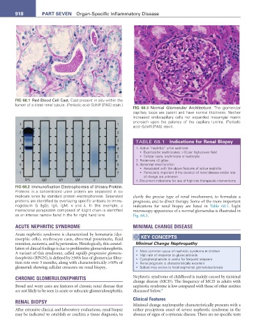
FIG 68.3 Normal Glomerular Architecture. The glomerular
capillary loops are patent and have normal thickness. Neither
increased endocapillary cells nor expanded mesangial matrix
encroach upon the patency of the capillary lumina. (Periodic
acid–Schiff [PAS] stain).
TABLE 68.1 Indications for renal Biopsy
1. Active “nephritic” urine sediment
• Dysmorphic erythrocytes: >10 per high-power field
• Cellular casts: erythrocyte or leukocyte
2. Proteinuria >2 g/day
3. Abnormal renal function
• Associated with the above features of active nephritis
• Particularly important if the duration of renal disease and/or rate
of change are unknown
63( ,J* ,J$ ,J0 κ λ 4. Document indications for use of high-risk therapeutic interventions
FIG 68.2 Immunofixation Electrophoresis of Urinary Protein.
Proteins in a concentrated urine protein are separated in six
replicate lanes by standard protein electrophoresis. Separated clarify the precise type of renal involvement, to formulate a
proteins are identified by overlaying specific antisera to immu- prognosis, and to direct therapy. Some of the more important
noglobulin G (IgG), IgA, IgM, κ and λ. In this example, a indications for renal biopsy are listed in Table 68.1. Light
monoclonal paraprotein composed of λlight chain is identified microscopy appearance of a normal glomerulus is illustrated in
as an intense narrow band in the far right hand lane. Fig. 68.3.
ACUTE NEPHRITIC SYNDROME MINIMAL CHANGE DISEASE
Acute nephritic syndrome is characterized by hematuria (dys-
morphic cells), erythrocyte casts, abnormal proteinuria, fluid KeY COnCePtS
retention, azotemia, and hypertension. Histologically, this constel- Minimal Change Nephropathy
lation of clinical findings is due to proliferative glomerulonephritis.
A variant of this syndrome, called rapidly progressive glomeru- • Most common cause of nephrotic syndrome in children
• High rate of response to glucocorticoids
lonephritis (RPGN), is defined by ≥50% loss of glomerular filtra- • Cyclophosphamide is useful for frequent relapsers
tion rate over 3 months, along with characteristically >50% of • Renal prognosis is characteristically excellent
glomeruli showing cellular crescents on renal biopsy.. • Subset may evolve to focal segmental glomerulosclerosis
CHRONIC GLOMERULONEPHRITIS Nephrotic syndrome of childhood is mainly caused by minimal
change disease (MCD). The frequency of MCD in adults with
Broad and waxy casts are features of chronic renal disease that nephrotic syndrome is low compared with those of other entities
are not likely to be seen in acute or subacute glomerulonephritis. discussed below. 4
Clinical Features
RENAL BIOPSY
Minimal change nephropathy characteristically presents with a
After extensive clinical and laboratory evaluations, renal biopsy rather precipitous onset of severe nephrotic syndrome in the
may be indicated to establish or confirm a tissue diagnosis, to absence of signs of a systemic disease. There are no specific tests