
Page 955 - Clinical Immunology_ Principles and Practice ( PDFDrive )
P. 955
922 Part Seven Organ-Specific Inflammatory Disease
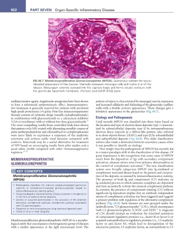
FIG 68.7 Membranoproliferative Glomerulonephritis (MPGN). Glomerulus exhibits the typical
lobulated appearance of this disease. Markedly increased mesangial cells and matrix in all of the
lobules. Mesangium extends outward into the capillary loops and forms double contours with
the glomerular basement membrane. (Periodic acid–Schiff [PAS] stain).
antihypertensive agents. Angiotensin antagonists have been shown pattern of injury is characterized by mesangial matrix expansion
to have a substantial antiproteinuric effect. Immunosuppres- and increased cellularity and thickening of the glomerular capillary
sive treatment is generally reserved for patients with persistent walls with a double contour appearance. These changes give a
high-grade proteinuria (>4 g/day. First-line immunosuppressive lobulated appearance to the glomerulus (Fig. 68.7).
therapy consists of cytotoxic drugs (usually cyclophosphamide)
in combination with glucocorticoids or a calcineurin inhibitor Etiology and Pathogenesis
16
(CSA or tacrolimus) with or without low-dose glucocorticoids. Until recently MPGN was classified into three types based on
The most compelling results from controlled trials have shown the location and type of electron dense deposits: type 1 character-
that patients with MN treated with alternating monthly courses of ized by subendothelial deposits; type II by intramembranous
pulse methylprednisolone and chlorambucil or cyclophosphamide electron dense deposits in a ribbon-like pattern (also referred
were more likely to experience a remission of the nephrotic to as dense deposit disease [DDD]); and type III by subendothelial
syndrome and achieve stable renal function compared with and subepithelial deposits (Fig. 68.8). This older classification
controls. Rituximab may be a useful alternative for treatment scheme also made a distinction between secondary causes when
of MN based on encouraging results from pilot studies and a it was possible to identify an etiology
good safety profile compared with other immunosuppressive New insight into the pathogenesis of MPGN has recently led
regimens. 16–19 to a major paradigm shift in the classification of this disease. Of
great importance is the recognition that some cases of MPGN
MEMBRANOPROLIFERATIVE result from the deposition of Igs with secondary complement
GLOMERULONEPHRITIS activation, whereas others arise from primary abnormalities in
20
the control of complement activation. The new classification
KeY COnCePtS system now broadly categorizes MPGN into Ig-mediated or
complement-mediated disease based on the pattern and composi-
Membranoproliferative Glomerulonephritis tion of the deposits, as assessed by immunofluorescence staining.
(MPGN) The presence of both Ig and complement (C3) indicates an
Ig-mediated process in which immune complexes are deposited
• Histologically classified into immune complex–mediated glomerulo- and then secondarily activate the classical complement pathway.
nephritis or complement-mediated glomerulonephritis based on
immunofluorescence staining pattern In contrast, the presence of complement staining (C3) without
• C3 glomerulopathy characterized by C3 accumulation in the glomeruli significant Ig deposition implies that an antibody-independent
in the form of electron-dense deposits. means of complement activation has been triggered and suggests
• Genetic or acquired abnormalities in the activation of the alternate a primary problem with regulation of the alternative complement
alternative complement pathway complement pathway associated pathway (Fig. 68.9). Such diseases are now grouped under the
with C3 glomerulopathy umbrella term, “C3 glomerulopathy” (C3G), which encompasses
• Response to immunosuppressive drug treatment generally poor both C3 glomerulonephritis (C3GN) and DDD. A diagnosis
21
• Tends to recur in renal allografts
of C3G should prompt an evaluation for inherited mutations
of complement regulatory proteins (i.e., factor H or factor I) or
Membranoproliferative glomerulonephritis (MPGN) is a morpho- acquired autoantibodies to regulatory proteins (i.e., C3 nephritic
logical entity that encompasses a heterogeneous group of diseases factor or anti-factor H), which lead to dysregulation of the
with a similar appearance at the light microscopic level. The alternative pathway. C3 nephritic factor, an autoantibody to C3