
Page 956 - Clinical Immunology_ Principles and Practice ( PDFDrive )
P. 956
CHaPter 68 Immunological Renal Diseases 923
$ %
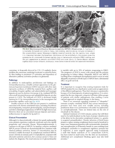
FIG 68.8 Membranoproliferative Glomerulonephritis (MPGN) Ultrastructure. A, Capillary wall
is markedly thickened and contains heavy, dark-staining, electron-dense immune complexes in
the subendothelial space. Mesangium (lighter material) extends into the capillary loop, where
it is interposed between the basement membrane and the endothelium; the process gives the
appearance of a massively thickened capillary loop on hematoxylin and eosin (H&E) staining, and
the split appearance by periodic acid–Schiff (PAS) and silver stains. B, Dense deposit disease;
capillary loops contain smooth, continuous, linear dense material within the basement membrane.
convertase, is frequently detected in C3G. C3 nephritic factor is variable with up to 30% of patients progressing to ESKD.
stabilizes the convertase, rendering it resistant to control by factor The prognosis of DDD is worse, with almost half the patients
H, thus leading to persistent C3 activation and deposition of progressing to kidney failure. Idiopathic MPGN and MPGN
alternative pathway activation products in glomeruli. resulting from complement dysregulation tend to recur in renal
allografts; recurrences of both types of MPGN have a detrimental
Pathology effect on graft survival.
In addition to endocapillary proliferation and findings on
immunofluorescence, a double contour appearance to the glo- Treatment
merular basement membrane (GBM) best seen with silver stain It is important to recognize that existing treatment trials for
represents synthesis of GBM-like material from capillary wall MPGN predate the new classification system and our appreciation
remodeling. Irregular capillary wall subendothelial and mesangial of the different pathogenesis. Thus their relevance is questionable
deposits are seen by electron microscopy in Ig- mediated MPGN; because of the heterogeneity of patient populations. It is antici-
often small intramembranous and subepithelial deposits are also pated that the new diagnostic categories will better define groups
present in C3GN. The hallmark of C3GN is dominant bright of patients with similar pathogenesis, thus enabling a more
staining for C3 by immunofluorescence in the mesangium and rational approach to targeted therapy in the future.
glomerular capillary walls (see Fig. 68.9). There is no consensus regarding treatment of “idiopathic”
Double contours of the GBM are also present in conditions immune complex–mediated MPGN, but various immunosup-
associated with chronic endothelial injury, including thrombotic pressants have been used with variable success, including
microangiopathy, transplant glomerulopathy, and preeclampsia, prednisone alone or in combination with either mycophenolate
and show a histological appearance of MPGN by light microscopy. or cyclophosphamide. There are limited data on the role of
However, there are no associated immune deposits and immu- rituximab in this disease.
nofluorescence is negative for Ig and C3. Optimal treatment of C3GN remains unclear but will likely
need to be individualized on the basis of proper identification
Clinical Presentation of the defect in the alternative complement system (i.e., genetic
Although it is characteristically a chronic low-grade nephropathy, vs acquired autoantibodies). Chronic infusions of fresh frozen
some patients experience nephrotic syndrome and even rapidly plasma to replace missing complement factors may be beneficial in
progressive and crescentic disease. Hypocomplementemia is some cases. Immunosuppression with corticosteroids, rituximab,
common in all types of MPGN. In immune complex-mediated and mycophenolate is of theoretic benefit in cases resulting from
MPGN, serum C4 levels are characteristically low, consistent with pathogenic antibodies to complement regulatory proteins but
classical pathway activation. Serum C3 concentration may be is unproven. A more targeted approach using eculizumab,
normal or mildly decreased. In complement-mediated MPGN, a mAb that blocks activation of C5 complement (C5b-9),
serum C4 levels are normal, and C3 is typically low, consistent with has been tested in small series, but the data are inconclusive.
alternative pathway activation, but a normal serum C3 does not Additional new complement blocking agents may also hold
exclude the diagnosis. The renal outcome in C3 glomerulopathy promise. 22