
Page 954 - Clinical Immunology_ Principles and Practice ( PDFDrive )
P. 954
CHaPter 68 Immunological Renal Diseases 921
$
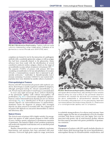
FIG 68.5 Membranous Nephropathy. Capillary walls are nearly
uniformly thickened but remain widely patent. Cellularity of the
glomerulus is normal (Periodic acid–Schiff [PAS] stain).
complexes are formed in situ by the interaction of a pathogenic
antibody with a constitutive glomerular antigen or with an antigen
that had been ectopically planted in the glomerulus. Major
advances have been made with the identification of several
constitutive podocyte surface antigens, which are the target of
autoantibodies in some subsets of patients with primary MN. 13,14
The majority (70-80%) of patients with primary MN have
circulating autoantibodies to M-type phospholipase A2 receptor
(PLA 2 R), a transmembrane receptor that is expressed in glo-
13
merular podocytes. Antibodies to thrombospondin type-1
domain containing 7A protein (THSDA7A) and neutral endo-
peptidase have been identified in smaller subsets of patients. 14
Clinicopathological Features
Patients with MN characteristically present with nephrotic syn-
drome. Renal biopsy is usually required to establish the diagnosis, %
although serological testing for relevant autoantibodies (i.e.,
anti–PLA 2 R) may be informative if renal biopsy is contraindicated. FIG 68.6 Membranous Nephropathy (Ultrastructure). A, Electron
In most cases, light microscopy shows uniform thickening micrograph demonstrates heavy, dark-staining immune complex
of the glomerular capillary walls without endocapillary cell deposits along the outer surface of the glomerular basement
proliferation (Fig. 68.5). Characteristic subepithelial (epimem- membrane and beneath the epithelial foot processes (hence
branous) and/or intramembranous deposits are seen on electron the terms subepithelial or epimembranous deposits). Note
microscopy (Fig. 68.6). Identification of PLA 2 R in glomerular the thickening and projections of the gray-staining basement
immune deposits (by immunofluorescence or immunohisto- membrane between the electron-dense deposits. B, Ultrastructure
chemistry) favors the diagnosis of primary MN; mesangial of a normal glomerular capillary wall for comparison.
deposits are often present in secondary MN. But a diagnosis of
secondary MN depends on concurrent abnormalities in clinical
and laboratory data.
is a relatively strong predictor of an adverse renal outcome. Some
Natural History studies suggest that the titer of autoantibody to PLA 2 R at baseline
The clinical course of primary MN is highly variable. On average, correlates with disease activity such that higher titers may be
about one-quarter of adult patients progress to ESKD within associated with greater risk of renal function decline, whereas
10 years. Another quarter experiences a spontaneous remission low titers may be associated with greater probability of spontane-
of proteinuria. The majority is likely to have persistent proteinuria ous remission. 15
and moderately impaired renal function over an extended period
of observation. Treatment
Baseline characteristics, such as severe nephrotic syndrome, Management of patients with MN usually includes diuretics to
hypertension, and azotemia, have been associated with poor reduce edema, lipid-lowering drugs (for severe hyperlipidemia),
outcomes. Protracted high-grade nephrotic-range proteinuria anticoagulant therapy for thromboembolic complications, and