
Page 986 - Clinical Immunology_ Principles and Practice ( PDFDrive )
P. 986
952 ParT SEvEN Organ-Specific Inflammatory Disease
A B
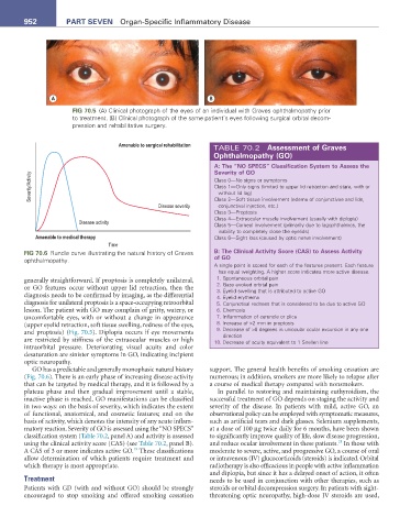
FIG 70.5 (A) Clinical photograph of the eyes of an individual with Graves ophthalmopathy prior
to treatment. (B) Clinical photograph of the same patient’s eyes following surgical orbital decom-
pression and rehabilitative surgery.
Amenable to surgical rehabilitation TABLE 70.2 assessment of Graves
Ophthalmopathy (GO)
a: The “NO SPECS” Classification System to assess the
Severity of GO
Severity/Activity Class 0—No signs or symptoms
Class 1—Only signs (limited to upper lid retraction and stare, with or
without lid lag)
conjunctival injection, etc.)
Disease severity Class 2—Soft tissue involvement (edema of conjunctivae and lids,
Class 3—Proptosis
Class 4—Extraocular muscle involvement (usually with diplopia)
Disease activity
Class 5—Corneal involvement (primarily due to lagophthalmos, the
inability to completely close the eyelids)
Amenable to medical therapy Class 6—Sight loss (caused by optic nerve involvement)
Time
FIG 70.6 Rundle curve illustrating the natural history of Graves B: The Clinical activity Score (CaS) to assess activity
ophthalmopathy. of GO
A single point is scored for each of the features present. Each feature
has equal weighting. A higher score indicates more active disease.
generally straightforward. If proptosis is completely unilateral, 1. Spontaneous orbital pain
or GO features occur without upper lid retraction, then the 2. Gaze evoked orbital pain
3. Eyelid swelling that is attributed to active GO
diagnosis needs to be confirmed by imaging, as the differential 4. Eyelid erythema
diagnosis for unilateral proptosis is a spaceoccupying retroorbital 5. Conjunctival redness that is considered to be due to active GO
lesion. The patient with GO may complain of gritty, watery, or 6. Chemosis
uncomfortable eyes, with or without a change in appearance 7. Inflammation of caruncle or plica
(upper eyelid retraction, soft tissue swelling, redness of the eyes, 8. Increase of >2 mm in proptosis
and proptosis) (Fig. 70.5). Diplopia occurs if eye movements 9. Decrease of >8 degrees in uniocular ocular excursion in any one
direction
are restricted by stiffness of the extraocular muscles or high 10. Decrease of acuity equivalent to 1 Snellen line
intraorbital pressure. Deteriorating visual acuity and color
desaturation are sinister symptoms in GO, indicating incipient
optic neuropathy.
GO has a predictable and generally monophasic natural history support. The general health benefits of smoking cessation are
(Fig. 70.6). There is an early phase of increasing disease activity numerous; in addition, smokers are more likely to relapse after
that can be targeted by medical therapy, and it is followed by a a course of medical therapy compared with nonsmokers.
plateau phase and then gradual improvement until a stable, In parallel to restoring and maintaining euthyroidism, the
inactive phase is reached. GO manifestations can be classified successful treatment of GO depends on staging the activity and
in two ways: on the basis of severity, which indicates the extent severity of the disease. In patients with mild, active GO, an
of functional, anatomical, and cosmetic features; and on the observational policy can be employed with symptomatic measures,
basis of activity, which denotes the intensity of any acute inflam such as artificial tears and dark glasses. Selenium supplements,
matory reaction. Severity of GO is assessed using the “NO SPECS” at a dose of 100 µg twice daily for 6 months, have been shown
classification system (Table 70.2, panel A) and activity is assessed to significantly improve quality of life, slow disease progression,
21
using the clinical activity score (CAS) (see Table 70.2, panel B). and reduce ocular involvement in these patients. In those with
19
A CAS of 3 or more indicates active GO. These classifications moderate to severe, active, and progressive GO, a course of oral
allow determination of which patients require treatment and or intravenous (IV) glucocorticoids (steroids) is indicated. Orbital
which therapy is most appropriate. radiotherapy is also efficacious in people with active inflammation
and diplopia, but since it has a delayed onset of action, it often
Treatment needs to be used in conjunction with other therapies, such as
Patients with GD (with and without GO) should be strongly steroids or orbital decompression surgery. In patients with sight
encouraged to stop smoking and offered smoking cessation threatening optic neuropathy, highdose IV steroids are used,