
Page 1127 - Hall et al (2015) Principles of Critical Care-McGraw-Hill
P. 1127
766 PART 6: Neurologic Disorders
junction testing. Peripheral motor nerve stimulation elicits a compound
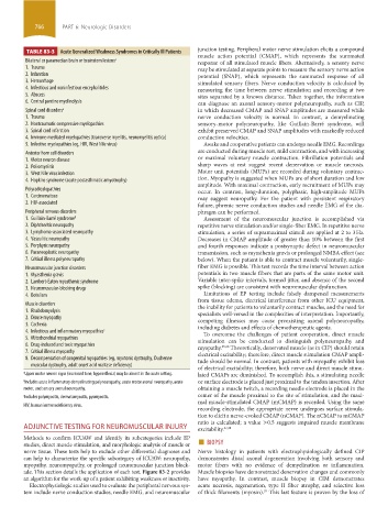
TABLE 83-3 Acute Generalized Weakness Syndromes in Critically Ill Patients
muscle action potential (CMAP), which represents the summated
Bilateral or paramedian brain or brainstem lesions a response of all stimulated muscle fibers. Alternatively, a sensory nerve
1. Trauma may be stimulated at separate points to measure the sensory nerve action
2. Infarction potential (SNAP), which represents the summated response of all
3. Hemorrhage stimulated sensory fibers. Nerve conduction velocity is calculated by
4. Infectious and noninfectious encephalitides measuring the time between nerve stimulation and recording at two
5. Abscess sites separated by a known distance. Taken together, the information
6. Central pontine myelinolysis can diagnose an axonal sensory-motor polyneuropathy, such as CIP,
Spinal cord disorders a in which decreased CMAP and SNAP amplitudes are measured while
1. Trauma nerve conduction velocity is normal. In contrast, a demyelinating
2. Nontraumatic compressive myelopathies sensory-motor polyneuropathy, like Guillain-Barré syndrome, will
3. Spinal cord infarction exhibit preserved CMAP and SNAP amplitudes with markedly reduced
4. Immune-mediated myelopathies (transverse myelitis, neuromyelitis optica) conduction velocities.
5. Infective myelopathies (eg, HIV, West Nile virus) Awake and cooperative patients can undergo needle EMG. Recordings
Anterior horn cell disorders are conducted during muscle rest, mild contraction, and with increasing
1. Motor neuron disease or maximal voluntary muscle contraction. Fibrillation potentials and
2. Poliomyelitis sharp waves at rest suggest recent denervation or muscle necrosis.
3. West Nile virus infection Motor unit potentials (MUPs) are recorded during voluntary contrac-
4. Hopkins syndrome (acute postasthmatic amyotrophy) tion. Myopathy is suggested when MUPs are of short duration and low
amplitude. With maximal contraction, early recruitment of MUPs may
Polyradiculopathies occur. In contrast, long-duration, polyphasic, high-amplitude MUPs
1. Carcinomatous may suggest neuropathy. For the patient with persistent respiratory
2. HIV-associated failure, phrenic nerve conduction studies and needle EMG of the dia-
Peripheral nervous disorders phragm can be performed.
1. Guillain-Barré syndrome b Assessment of the neuromuscular junction is accomplished via
2. Diphtheritic neuropathy repetitive nerve stimulation and/or single-fiber EMG. In repetitive nerve
3. Lymphoma-associated neuropathy stimulation, a series of supramaximal stimuli are applied at 2 to 3 Hz.
4. Vasculitic neuropathy Decreases in CMAP amplitude of greater than 10% between the first
5. Porphyric neuropathy and fourth responses indicate a postsynaptic defect in neuromuscular
6. Paraneoplastic neuropathy transmission, such as myasthenia gravis or prolonged NMBA effect (see
7. Critical illness polyneuropathy below). When the patient is able to contract muscle voluntarily, single-
Neuromuscular junction disorders fiber EMG is possible. This test records the time interval between action
1. Myasthenia gravis potentials in two muscle fibers that are parts of the same motor unit.
2. Lambert-Eaton myasthenic syndrome Variable inter-spike intervals, termed jitter, and absence of the second
3. Neuromuscular-blocking drugs spike (blocking) are consistent with neuromuscular dysfunction.
4. Botulism Limitations of EP testing include falsely dampened measurements
from tissue edema, electrical interference from other ICU equipment,
Muscle disorders the inability for patients to voluntarily contract muscles, and the need for
1. Rhabdomyolysis specialists well-versed in the complexities of interpretation. Importantly,
2. Disuse myopathy competing illnesses may cause preexisting axonal polyneuropathy,
3. Cachexia including diabetes and effects of chemotherapeutic agents.
4. Infectious and inflammatory myopathies c To overcome the challenges of patient cooperation, direct muscle
5. Mitochondrial myopathies stimulation can be conducted to distinguish polyneuropathy and
6. Drug-induced and toxic myopathies myopathy. 25,26 Theoretically, denervated muscle (as in CIP) should retain
7. Critical illness myopathy electrical excitability; therefore, direct muscle stimulation CMAP ampli-
8. Decompensation of congenital myopathies (eg, myotonic dystrophy, Duchenne tude should be normal. In contrast, patients with myopathy exhibit loss
muscular dystrophy, adult onset acid maltase deficiency)
of electrical excitability; therefore, both nerve and direct muscle stimu-
a Upper motor neuron signs (increased tone, hyperreflexia) may be absent in the acute setting. lated CMAPs are diminished. To accomplish this, a stimulating needle
b Includes acute inflammatory demyelinating polyneuropathy, acute motor axonal neuropathy, acute or surface electrode is placed just proximal to the tendon insertion. After
motor, and sensory axonal neuropathy. obtaining a muscle twitch, a recording needle electrode is placed in the
c Includes polymyositis, dermatomyositis, pyomyositis. center of the muscle proximal to the site of stimulation, and the maxi-
mal muscle-stimulated CMAP (mCMAP) is recorded. Using the same
HIV, human immunodeficiency virus.
recording electrode, the appropriate nerve undergoes surface stimula-
tion to elicit a nerve-evoked CMAP (nCMAP). The nCMAP to mCMAP
ratio is calculated; a value >0.5 suggests impaired muscle membrane
ADJUNCTIVE TESTING FOR NEUROMUSCULAR INJURY excitability. 27,28
studies, direct muscle stimulation, and morphologic analysis of muscle or ■ BIOPSY
Methods to confirm ICUAW and identify its subcategories include EP
nerve tissue. These tests help to exclude other differential diagnoses and Nerve histology in patients with electrophysiologically defined CIP
can help to characterize the specific subcategory of ICUAW: neuropathy, demonstrates distal axonal degeneration involving both sensory and
myopathy, neuromyopathy, or prolonged neuromuscular junction block- motor fibers with no evidence of demyelination or inflammation.
ade. This section details the application of each test. Figure 83-2 provides Muscle biopsies have demonstrated denervation changes and commonly
an algorithm for the work-up of a patient exhibiting weakness or inactivity. have myopathy. In contrast, muscle biopsy in CIM demonstrates
Electrophysiologic studies used to evaluate the peripheral nervous sys- acute necrosis, regeneration, type II fiber atrophy, and selective loss
tem include nerve conduction studies, needle EMG, and neuromuscular of thick filaments (myosin). This last feature is proven by the loss of
29
section06.indd 766 1/23/2015 12:55:30 PM