
Page 1155 - Hall et al (2015) Principles of Critical Care-McGraw-Hill
P. 1155
794 PART 6: Neurologic Disorders
of CPP within a targeted range of 50 to 70 mm Hg can therefore be an compartmental parenchymal shifts in response to trajectories of increas-
important therapeutic strategy in providing a margin of “reserve” with ing pressure differentials. The end result of untreated, progressive brain
which the brain can compensate for challenges of normal perfusion via edema is herniation and ultimately brain death. Despite the obvious
fluctuations in ICP or MAP. clinical importance of cerebral edema, the precise mechanisms of water
As pressure autoregulation and microcirculatory homeostasis may transport and accumulation of excess water within the brain remain
be severely disrupted in the brain-injured patient, ischemia can result unclear. A series of recent studies on cerebral edema focused on the
even in the presence of adequate ICP and CPP. Adjusting the target CPP glial water protein channel aquaporin-4 (AQP4), among others, such as
in a particular patient based on the clinical situation, the underlying AQP1 and AQP9, that have been shown to facilitate astrocyte swelling
etiology of brain injury and the vasoreactive state is therefore necessary. (“cytotoxic edema”) and also to be responsible for the reabsorption
Some studies support the concept of CPP targeting based on cerebral of extracellular edema fluid (“vasogenic edema”). Therefore, AQP4
vasoreactivity monitoring. 11,12 In addition, improved tools for ICP mea- modulation via pharmacologic interventions has become an interesting
surements (ie, via minimal invasive intraparenchymal devices) with potential therapeutic approach. 14-16 AQP4 knock-out, or disruption of
continuous CPP determinations and the ability to correlate additional its polarized expression pattern, mitigates brain water accumulation and
brain monitoring parameters such as cerebral blood flow, oxygenation, therefore decreases associated ischemia, water intoxication, and hypona-
and chemical profiling allow multimodal, real-time pathophysiologic tremia in animal models. 17
analysis of brain injury at the bedside. The most common types of cerebral edema are cytotoxic edema from
■ PLATEAU WAVES cellular injury and swelling, and vasogenic edema from breakdown
of the blood-brain barrier and interstitial fluid extravasation. Other
One of the most feared complications of intracranial hypertension and types, such as hydrocephalic edema, ischemic edema (a combination
poor intracranial compliance is the development of plateau waves (PW) of cytotoxic and vasogenic edema), osmotic edema, and hydrostatic
(Fig. 86-4). These waves are associated with acute elevations in ICP or interstitial edema have also been characterized as distinct entities
ranging from 50 to 100 mm Hg. They typically occur in patients with based on their underlying mechanisms and the predominant location
reduced intracranial compliance discussed later. Plateau waves can last of fluid. 18-20 Table 86-3 lists the categories of cerebral edema along with
from several minutes to more extended durations in severe cases and their distinguishing characteristics. Clinically, vasogenic and cytotoxic
are rapid in onset and offset. While there are many causes of PW, one edema are most frequently encountered. Disruption of the blood-brain
important and common mechanism is generalized cerebral vasodilation barrier results in plasma-derived, protein-rich exudate accumulating in
from an uncontrolled autoregulatory response to a decrease in systemic the extracellular white matter, constituting vasogenic edema. Despite
blood pressure. Other causes include processes that increase CBF and the commonly encountered severity of vasogenic edema, CBF is often
13
CBV (Table 86-2). Since compromised CPP can play an important role unaffected and cellular mechanisms remain intact. Among the disease
in the occurrence of the most severe PW, relative CPP drops should be entities with predominant but variable degrees of vasogenic edema are
avoided and/or rapidly treated. Similarly, during a PW, maneuvers that brain tumors (Fig. 86-10), abscesses, traumatic brain injury, and menin-
aim to correct CPP toward the target range, such as swift blood pressure gitis. Corticosteroids play a primary role in reducing this type of edema,
augmentation, will potentially abort the PW in many circumstances. and their effect is most profound when vasogenicity is the primary
21
Even if blood pressure augmentation does not abort the process, it will etiology, as with brain tumors, and to a lesser degree with abscesses.
likely reduce cerebral ischemia until other treatment modalities can suc- In comparison, osmotic agents have little beneficial effect on vasogenic
18
cessfully lower the uncontrolled ICP. edema. Cytotoxic edema, in contrast, is characterized by intracellular
swelling of neurons, glia, and endothelial cells with an accompanying
reduction in the extracellular space. It occurs without disruption of the
CEREBRAL EDEMA, MASS EFFECT, BRAIN HERNIATION blood-brain barrier, and is primarily due to cellular energy depletion,
■ CEREBRAL EDEMA AND MASS EFFECT which results in failure of the ATP-dependent sodium pump and accu-
mulation of sodium and water within cells. Cytotoxic edema can occur
18
Cerebral edema is an increase in tissue water content within and or in both gray and white matter. Hypoperfusion (ischemic) injuries are
around brain cells. Patients with acute brain injuries invariably present most classically associated with cytotoxic edema. While edema in TBI
with different degrees of edema as a result of different mechanisms of was thought to be vasogenic in origin, clinical and experimental studies
intra-and extraaxial injury. The consequences of uncontrolled edema indicate that cytotoxic edema predominates following TBI. 22-24 This may
range from cerebral ischemia to mechanical compression of brain explain why drugs that attenuate vasogenic brain edema (eg, corticoste-
tissue. Initially, edema affects a regional area and can progress to roids) are only beneficial in certain conditions (eg, tumors) but not in
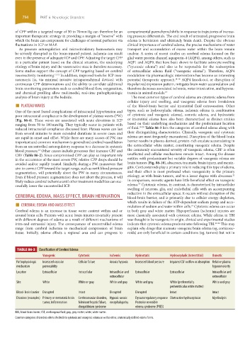
TABLE 86-3 Classification of Cerebral Edema
Vasogenic Cytotoxic Ischemic Hydrostatic Hydrocephalic (Interstitial) Osmotic
Pathophysiologic Increased vascular Cellular failure Anoxia/hypoxia Increased blood pressure Impaired CSF outflow or absorption Relative plasma
mechanism permeability hypoosmolarity
Location Extracellular Intracellular Intracellular and Extracellular Extracellular Intracellular and
extracellular extracellular
Site White White or gray White and gray White and gray White (preferentially White and gray
periventricular white matter)
Blood- brain barrier Disrupted Intact Disrupted Disrupted Intact Intact
Disorders (examples) Primary or metastatic brain Cerebrovascular disorders, Hypoxic-anoxic Dysautoregulatory response Obstructive hydrocephalus Myelinolysis
tumor, Inflammation fulminant hepatic failure, encephalopathy Posterior reversible
disequilibrium syndrome edema syndrome (PRES)
BBB, blood-brain barrier; CSF, cerebrospinal fluid; gray, gray matter; white, white matter.
Common categories of cerebral edema divided into cytotoxic and vasogenic edema as well as other, anatomically defined edema forms.
section06.indd 794 1/23/2015 12:55:51 PM