
Page 1186 - Hall et al (2015) Principles of Critical Care-McGraw-Hill
P. 1186
CHAPTER 87: Neuromuscular Diseases Leading to Respiratory Failure 823
and a VC <15 to 20 mL/kg greatly increases the likelihood of respiratory MIP less negative than −46 cm H O (<4.5 kPa; 1 kPa = 10.19 cm H O),
2
2
failure. 2,5,12 sleep-disordered breathing is common. 17
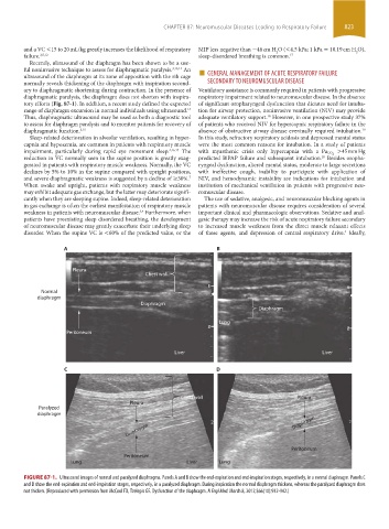
Recently, ultrasound of the diaphragm has been shown to be a use-
ultrasound of the diaphragm at its zone of apposition with the rib cage ■ GENERAL MANAGEMENT OF ACUTE RESPIRATORY FAILURE
An
ful noninvasive technique to assess for diaphragmatic paralysis.
3,13,14
normally reveals thickening of the diaphragm with inspiration second- SECONDARY TO NEUROMUSCULAR DISEASE
ary to diaphragmatic shortening during contraction. In the presence of Ventilatory assistance is commonly required in patients with progressive
diaphragmatic paralysis, the diaphragm does not shorten with inspira- respiratory impairment related to neuromuscular disease. In the absence
tory efforts (Fig. 87-1). In addition, a recent study defined the expected of significant oropharyngeal dysfunction that dictates need for intuba-
range of diaphragm excursion in normal individuals using ultrasound. tion for airway protection, noninvasive ventilation (NIV) may provide
14
Thus, diaphragmatic ultrasound may be used as both a diagnostic tool adequate ventilatory support. However, in one prospective study 37%
18
to assess for diaphragm paralysis and to monitor patients for recovery of of patients who received NIV for hypercapnic respiratory failure in the
19
diaphragmatic function. 3,15 absence of obstructive airway disease eventually required intubation.
Sleep-related deterioration in alveolar ventilation, resulting in hyper- In this study, refractory respiratory acidosis and depressed mental status
capnia and hypoxemia, are common in patients with respiratory muscle were the most common reasons for intubation. In a study of patients
impairment, particularly during rapid eye movement sleep. 4,16,17 The with myasthenic crisis only hypercapnia with a Pa CO 2 >45 mm Hg
reduction in VC normally seen in the supine position is greatly exag- predicted BiPAP failure and subsequent intubation. Besides oropha-
20
gerated in patients with respiratory muscle weakness. Normally, the VC ryngeal dysfunction, altered mental status, moderate to large secretions
declines by 5% to 10% in the supine compared with upright positions, with ineffective cough, inability to participate with application of
and severe diaphragmatic weakness is suggested by a decline of ≥30%. NIV, and hemodynamic instability are indications for intubation and
7
When awake and upright, patients with respiratory muscle weakness institution of mechanical ventilation in patients with progressive neu-
may exhibit adequate gas exchange, but the latter may deteriorate signifi- romuscular disease.
cantly when they are sleeping supine. Indeed, sleep-related deterioration The use of sedative, analgesic, and neuromuscular blocking agents in
in gas exchange is often the earliest manifestation of respiratory muscle patients with neuromuscular disease requires consideration of several
weakness in patients with neuromuscular disease. Furthermore, when important clinical and pharmacologic observations. Sedative and anal-
3,4
patients have preexisting sleep disordered breathing, the development gesic therapy may increase the risk of acute respiratory failure secondary
of neuromuscular disease may greatly exacerbate their underlying sleep to increased muscle weakness from the direct muscle relaxant effects
disorder. When the supine VC is <60% of the predicted value, or the of these agents, and depression of central respiratory drive. Ideally,
4
A B
Pleura
Chest wall
Normal
diaphragm
Diaphragm
Diaphragm
Lung
Peritoneum
Liver Liver
C D
Chest wall Pleura
Pleura
Paralyzed
diaphragm
Diaphragm Diaphragm
Peritoneum
Peritoneum
Lung Liver Lung
FIGURE 87-1. Ultrasound images of normal and paralyzed diaphragms. Panels A and B show the end-expiration and end-inspiration stages, respectively, in a normal diaphragm. Panels C
and D show the end-expiration and end-inspiration stages, respectively, in a paralyzed diaphragm. During inspiration the normal diaphragm thickens, whereas the paralyzed diaphragm does
not thicken. (Reproduced with permission from McCool FD, Tzelepis GE. Dysfunction of the diaphragm. N Engl Med. March 8, 2012;366(10):932-942.)
section06.indd 823 1/23/2015 12:56:18 PM