
Page 1182 - Hall et al (2015) Principles of Critical Care-McGraw-Hill
P. 1182
CHAPTER 86: Intracranial Pressure: Monitoring and Management 819
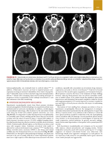
FIGURE 86-21. Global ischemia and cerebral edema. Unenhanced head CT of an 83-year-old man who complained of sudden severe headache during dialysis, arrested and was resus-
citated for 2 hours. Global brain swelling described as no distinction of gray and white matter and effacement of all gyri and sulci can be identified. Subarachnoid hemorrhages secondary to
ruptured aneurysm with mild intraventricular extension, which lead to hydrocephalus, are also shown.
leukoencephalopathy, can eventually lead to cerebral edema. 200,201 In conditions, especially with concomitant use of cytotoxic drugs, immuno-
205
addition, inflammatory responses can lead to impaired pressure auto- suppressives, and acute or chronic renal diseases. Treatment is focused
regulation resulting in increased vulnerability of the brain to hypoperfu- on removal of the causative agent, oxygenation, and treatment of seizures.
202
sion. Ultimately, sepsis survivors may have long-term neuropsychiatric Blood pressure control is the focus of the treatment of brain swelling;
deficits. Patients with underlying sepsis and fluctuating sensorium or however, reducing blood pressure does not achieve immediate resolu-
199
progressive decline in arousability should undergo noncontrast CT scan tion of cerebral edema. If the patient already has significant intracranial
and continuous video EEG monitoring. hypertension and diminished intracranial compliance (aided by clinical
■ HYPERTENSIVE ENCEPHALOPATHY AND ECLAMPSIA and imaging parameters), ICP monitoring may be necessary to guide the
pace and degree of blood pressure reduction with respect to maintaining
Hypertensive encephalopathy results from blood pressure elevations adequate CPP.
beyond autoregulatory thresholds leading to increased extracellular water, Mechanisms of brain swelling from eclampsia are similar to those in
predominantly by hydrostatic mechanisms. In addition, variable degrees hypertensive encephalopathy. However, the management of eclampsia-
of parenchymal hemorrhage, often localized in the end-arterial border associated intracranial hypertension has the added priority of urgent fetal
zones along the frontal and posterior parietal convexities, can occur delivery. Cesarean section is the preferred mode of delivery in almost all
(Fig. 86-22A). It is important to realize that hypertensive encephalopathy cases. Spinal anesthesia should be avoided due to the risk of precipitating
is a reversible cause of brain swelling and the extent does not necessarily central herniation with CSF drainage. General anesthesia should include
correlate with the extent of neuronal injury. A typical MRI pattern on close attention to the blood pressure to avoid degrees of lowering that
fluid-attenuated inversion recovery (FLAIR)–weighted imaging identi- could compromise cerebral perfusion. In general, successful manage-
fies what has also been described as posterior reversible edema syndrome ment of intracranial hypertension is best guided with a parenchymal ICP
(PRES). Predominantly affected regions are the bilateral parietooccipital monitor.
areas with vasogenic edema. Sometimes subcortical white matter is Both hypertensive encephalopathy and eclampsia can be associated
affected, but cortical involvement is also common. 125,203,204 Another term, with ominous clinical presentations and imaging studies. Neither
diffuse reversible edema syndrome (DRES) is also being used instead midposition unreactive pupils with extensor posturing nor CT changes
of PRES since the frontoparietal lobes are often involved (Fig. 86-22B). suggestive of bilateral end-arterial border zone infarctions with
PRES/DRES are descriptive clinicoradiological findings, and physicians hemorrhage should deter aggressive management in such patients. In
involved in the care of acutely, difficult-to-control hypertensive patients our experience, both scenarios can potentially lead to good outcomes
must have a high suspicion of the clinical spectrum associated with these when treated promptly and aggressively.
section06.indd 819 1/23/2015 12:56:15 PM