
Page 1185 - Hall et al (2015) Principles of Critical Care-McGraw-Hill
P. 1185
822 PART 6: Neurologic Disorders
accessory muscles of inspiration and paradoxical inward movement of
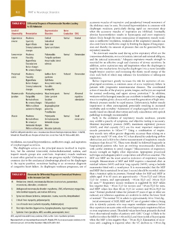
TABLE 87-2 Differential Diagnosis of Neuromuscular Disorders leading
to ICU Admission the abdomen may be seen. Nocturnal hypoventilation is common with
diaphragm weakness, particularly during rapid eye movement sleep
level of Representative Nerve when the accessory muscles of inspiration are inhibited. Eventually,
Abnormality Presentation Disorders Conduction EMG alveolar hypoventilation results in hypercapnia and overt respiratory
Upper motor Weakness Cortical Normal Normal failure. Even though the main consequence of inspiratory muscle weak-
neuron Spasticity Subcortical ness is ineffective ventilation, weakness of the inspiratory muscles can
Hyperreflexia Brain stem also contribute to ineffective cough by limiting the degree of lung expan-
Sensory/autonomic Spinal cord lesions sion and thereby the amount of pressure that can be generated by the
changes expiratory muscles.
The dominant muscles used during active expiratory effort are the
Lower motor Weakness Poliomyelitis Normal Denervation transversus abdominis, rectus abdominis, internal and external obliques,
neuron Flaccidity Postpolio syndrome and the internal intercostals. Adequate expiratory muscle strength is
2
Hyporeflexia Amyotrophic lateral essential for an effective cough and clearance of airway secretions. In
Fasciculations sclerosis addition, active expiration may aid inspiration when the diaphragm is
Bulbar changes weak by two mechanisms: forcing the diaphragm into a more favorable
No sensory changes
length-tension position and increasing the elastic recoil energy of the
Peripheral Weakness Guillian-Barre Reduced Denervation chest wall, both of which may enhance the forcefulness of subsequent
nerve Flaccidity syndrome inspiration.
Hyporeflexia Diphtheria Bulbar impairment greatly increases the risk for aspiration of oro-
Bulbar changes Heavy metal toxicity pharyngeal secretions, a common cause of acute respiratory failure in
Sensory/autonomic Vasculitic patients with progressive neuromuscular diseases. The coordinated
changes neuropathy action of muscles of the pharynx, palate, tongue, and larynx are required
Neuromuscular Fluctuating weakness Myasthenia gravis Normal Abnormal for normal swallowing and upper airway protection. In addition,
2,5
junction Fatigability Eaton-Lambert repetitive weakness of the laryngeal muscles can contribute to ineffective coughing
Normal reflexes Botulism stimulation since incomplete glottic closure will prevent the generation of high intra-
No sensory changes Tick paralysis thoracic pressure needed to expel mucus. Unfortunately, bulbar muscle
With or without Organophosphate impairment is often unrecognized, potentially resulting in increased
5
autonomic changes poisoning morbidity and mortality. Assessment of oropharyngeal function is pri-
Penicillamine marily based on clinical observation and early consultation with speech
pathology is strongly recommended.
Muscle Weakness Polymyositis Normal Small
Early in the evolution of respiratory muscle weakness, patients
Normal reflexes Dermatomyositis motor units may exhibit a paucity of symptoms, and objective testing is necessary.
No sensory or Metabolic myopa-
Autonomic changes thies Maximal inspiratory pressure (MIP), maximum expiratory pressure
(MEP), and vital capacity (VC) are the most important respiratory
With or without pain Muscular dystrophy
muscle parameters to follow. 2,3,6,7 Using a combination of respira-
Modified with permission from Luce J. Neuromuscular diseases leading to respiratory failure. In: Hall JB, tory muscle tests offers greater diagnostic accuracy than relying on a
Schmidt GA, Wood LDH, eds. Principles of Critical Care. 3rd ed. New York, NY: McGraw-Hill; 2005.
single test result. Of note, slow VC is less altered by underlying airflow
8
obstruction and is felt to be a better measurement of respiratory muscle
ventilation, nocturnal hypoventilation, ineffective cough, and aspiration weakness than forced VC. These tests should be followed frequently in
of oropharyngeal secretions. hospitalized patients who have an evolving neuromuscular disorder,
The diaphragm serves as the principal muscle involved in inspira- with careful attention to serial changes. Measurements of respiratory
9
tion, but the external intercostal, sternocleidomastoid, scalene, and muscle strength are highly effort dependent. Appropriate procedural
trapezii muscle groups also contribute. Inspiratory muscle weakness technique and adequate patient cooperation and effort are essential. The
2
is most often gradual in onset, but can progress rapidly. Orthopnea is MIP and MEP are the most sensitive indicators of respiratory muscle
common due to the mechanical disadvantage placed on the diaphragm strength. Measurement of MIP and MEP requires a maximal effort at
in the supine position, sometimes leading to an erroneous diagnosis residual volume (MIP) and total lung capacity (MEP), using a bedside
of congestive heart failure. As diaphragm weakness progresses, use of manometer fitted with a mouthpiece. It is recommended that the MIP
and MEP that is sustained for at least 1 second should be recorded rather
TABLE 87-3 Mnemonic for Differential Diagnosis of Generalized Weakness than a transient spike in pressure. Normal values for MIP and MEP in
in the Intensive Care Unit adults aged 18 to 65 years are approximately −70 cm H O and 100 cm
2
H O for women, and approximately −95 cm H O and 140 cm H O
2
2
2
M Medications: steroids, neuromuscular blockers (cisatracurium, pancuronium, for men. Respiratory muscle weakness is suggested by MIP values
6,10
vecuronium), zidovudine, amiodarone less negative than −30 cm H O for women and −45 cm H O for men,
2
2
U Undiagnosed neuromuscular disorder: myasthenia, LEMS, inflammatory myopathies, and MEP values less than 60 cm H O for women and 80 cm H O for
2
2
8
mitochondrial myopathy, acid maltase deficiency men. Normal predicted values for patients 65 years of age or older are
reduced, and reference equations are available to define the lower limit
S Spinal cord disease (ischemia, compression, trauma, vasculitis, demyelination)
of normal. The normal VC in adults is approximately 50 to 70 mL/kg.
11
C Critical illness myopathy, polyneuropathy Serial assessment of MIP, MEP, and VC are of greatest value in being
L Loss of muscle mass (cachectic myopathy, rhabdomyolysis) able to identify patients who may require ventilator assistance before
E Electrolyte disorders (hypokalemia, hypophosphatemia, hypermagnesemia) they experience an acute crisis with overt hypercapnic respiratory failure
or even respiratory arrest. Threshold values have been primarily derived
S Systemic illness (porphyria, AIDS, vasculitis, paraneoplastic, toxic)
from observational studies of patients with GBS. Cough is likely to be
9
AIDS, acquired immunodeficiency syndrome; LEMS, Lamber-Eaton myasthenic syndrome. ineffective when the MEP is <40 cm H O, and there is risk of hypercapnia
2
Reproduced with permission from Maramatton BV, Wijdicks EFM. Acute neuromuscular weakness in the when the MIP is less negative than −30 cm H O. Elimination of secre-
2
intensive care unit. Crit Care Med. November 2006;34(11):2835-2841. tions with coughing is impaired when the VC declines to <30 mL/kg
section06.indd 822 1/23/2015 12:56:17 PM