
Page 1240 - Hall et al (2015) Principles of Critical Care-McGraw-Hill
P. 1240
CHAPTER 90: Bleeding Disorders 847
TABLE 90-3 Essential Events of Coagulation Cascade
Step Consequence Direct Mediator(s) Mechanism
1. Activation of prothrombin to Conversion of fibrinogen to fibrin Activated factor V Tissue factor and calcium bind to factor VII, resulting in activation.
thrombin Activated VII activates factor X, factor Xa activates prothrombin
2. Conversion of fibrinogen Formation of insoluble fibrin multimers Thrombin Activation of prothrombin to thrombin
to fibrin and cross-linking/aggregation of platelets
3. Propagation and amplification Rapid burst of fibrin formation Thrombin Thrombin itself activates factor V to Va, factor VII to VIIIa and
of thrombin production mediates conversion of XI to XIa
4. Termination of thrombin Cessation of fibrin formation Antithrombin, Thrombomodulin, Antithrombin neutralizes thrombin, Xa, IXa, XIIa, and XIa.
activation and removal of active protein C, protein S, tissue factor Thrombomodulin binds thrombin to inhibit platelet activation
thrombin pathway inhibitor (TFPI) and cleavage of fibrinogen. Proteins C and S inactivate factor Va
and VIIIa. TFPI inhibits Xa
5. Fibrinolysis Cleavage of polymerized fibrin and release Plasmin, tissue-type plasminogen Plasminogen and tissue plasminogen activator form complex
of fibrin degradation products activator, urokinase with fibrin leading to active proteolytic plasmin
understanding of common laboratory tests including prothrombin time coagulation), severe inflammatory states (eg, sepsis, trauma), and inhi-
and activated partial thromboplastin time (see “Laboratory Testing of bition of activity in autoimmune disease (eg, antibody inhibitors).
Coagulation Function” below). 27
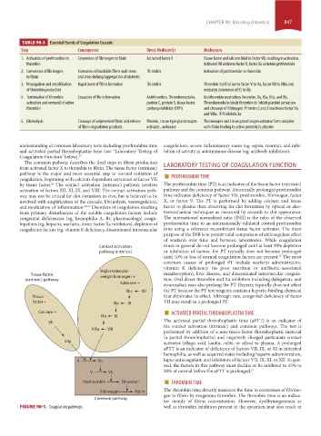
The common pathway describes the final steps in fibrin production
from activated factor X to thrombin to fibrin. The tissue factor (extrinsic) LABORATORY TESTING OF COAGULATION FUNCTION
pathway is the major and most essential step in normal initiation of ■
coagulation, beginning with calcium-dependent activation of factor VII PROTHROMBIN TIME
by tissue factor. The contact activation (intrinsic) pathway involves The prothrombin time (PT) is an indicator of the tissue factor (extrinsic)
28
activation of factors XII, XI, IX, and VIII. The contact activation path- pathway and the common pathway. Abnormally prolonged prothrombin
way may not be critical for clot formation in vivo, but is believed to be time indicates deficiency of factor VII, prothrombin, fibrinogen, factor
involved with amplification of the cascade, fibrinolysis, vasoregulation, X, or factor V. The PT is performed by adding calcium and tissue
and modulation of inflammation. 29-31 Disorders of coagulation resulting factor to plasma then observing for clot formation by optical or elec-
from primary disturbances of the soluble coagulation factors include tromechanical techniques as measured by seconds to clot appearance.
congenital deficiencies (eg, hemophilia A, B), pharmacologic coagu- The international normalized ratio (INR) is the ratio of the observed
lopathies (eg, heparin, warfarin, direct factor Xa inhibitors), depletion of prothrombin time to an internationally validated control prothrombin
coagulation factors (eg, vitamin K deficiency, disseminated intravascular time using a reference recombinant tissue factor activator. The main
purpose of the INR is to permit valid comparison of anticoagulant effect
of warfarin over time and between laboratories. While coagulation
Contact activation times in general do not become prolonged until at least 50% depletion
pathway (intrinsic) or inhibition of factors, the PT typically does not become prolonged
until 10% or less of normal coagulation factors are present. The most
27
common causes of prolonged PT include warfarin administration,
vitamin K deficiency (in poor nutrition or antibiotic-associated
High-molecular-
Tissue factor weight kininogen + malabsorption), liver disease, and disseminated intravascular coagula-
(extrinsic) pathway tion. Oral direct thrombin and Xa inhibitors including dabigatran, and
Kallikrein + rivaroxaban may also prolong the PT. Heparin typically does not affect
XIIa XII the PT because the PT test reagents contain a heparin-binding chemical
Tissue that eliminates its effect. Although rare, congenital deficiency of factor
factor + XIa XI VII may result in a prolonged PT.
Calcium + ■ ACTIVATED PARTIAL THROMBOPLASTIN TIME
IXa IX
Vll The activated partial thromboplastin time (aPTT) is an indicator of
the contact activation (intrinsic) and common pathways. The test is
Vllla Vlll
performed by addition of a non-tissue-factor thromboplastic material
(a partial thromboplastin) and negatively charged particulate contact
Vlla activator (ellagic acid, kaolin, celite, or silica) to plasma. A prolonged
aPTT is an indicator of deficiency of factors VII, IX, or XI in inherited
hemophilia, as well as acquired states including heparin administration,
X Xa lupus anticoagulant, and inhibitors of factors VII, IX, XI, or XII. In gen-
eral, the factors in this pathway must decline or be inhibited to 15% to
V Va 30% of normal before the aPTT is prolonged. 27
■
Prothrombin Thrombin THROMBIN TIME
The thrombin time directly measures the time to conversion of fibrino-
Fibrinogen Fibrin
gen to fibrin by exogenous thrombin. The thrombin time is an indica-
Common pathway
tor mainly of fibrin concentration. However, dysfibrinogenemia as
FIGURE 90-1. Coagulation pathways. well as thrombin inhibitors present in the specimen may also result in
section07.indd 847 1/21/2015 7:42:42 AM