
Page 1241 - Hall et al (2015) Principles of Critical Care-McGraw-Hill
P. 1241
848 PART 7: Hematologic and Oncologic Disorders
prolonged thrombin time (eg, heparin, bivalirudin, argatroban, high
concentrations of serum proteins in multiple myeloma). A similar test, Torsion force
the reptilase time (RT), uses snake venom enzyme similar to thrombin measurement
which cleaves fibrinogen but is not sensitive to heparin and thus distin-
guishes hypofibrinogenemia from heparin anticoagulation.
■ ACTIVATED WHOLE BLOOD CLOTTING TIME
The activated clotting time (ACT) is performed by adding negatively
charged particles (celite, kaolin) to a sample of freshly drawn whole
blood and measuring the formation of clot. The ACT is influenced
by both the soluble coagulation factors as well as the platelets present
in blood sample. Although the test closely resembles the aPTT, it is
clinically less sensitive to the effects of heparin and hence is used for
monitoring coagulation in the setting of high heparin infusions such as
coronary artery bypass surgery, percutaneous vascular procedures, and
extracorporeal membrane oxygenation.
■ FIBRIN DEGRADATION PRODUCTS AND D-DIMER
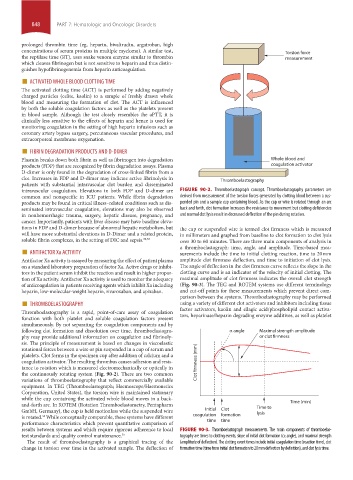
Plasmin breaks down both fibrin as well as fibrinogen into degradation Whole blood and
products (FDP) that are recognized by fibrin degradation assays. Plasma coagulation activator
D-dimer is only found in the degradation of cross-linked fibrin from a
clot. Increases in FDP and D-dimer may indicate active fibrinolysis in Thromboelastography
patients with substantial intravascular clot burden and disseminated
intravascular coagulation. Elevations in both FDP and D-dimer are FIGURE 90-2. Thromboelastograph concept. Thromboelastography parameters are
common and nonspecific in ICU patients. While fibrin degradation derived from measurement of the torsion forces generated by clotting blood between a sus-
products may be found in critical illness–related conditions such as dis- pended pin and a sample cup containing blood. As the cup or wire is rotated through an arc
seminated intravascular coagulation, elevations may also be observed back and forth, clot formation increases the resistance to movement but clotting deficiencies
in nonhemorrhagic trauma, surgery, hepatic disease, pregnancy, and and normal clot lysis result in decreased deflection of the pin during rotation.
cancer. Importantly, patients with liver disease may have baseline eleva-
tions in FDP and D-dimer because of abnormal hepatic metabolism, but the cup or suspended wire is termed clot firmness which is measured
will have more substantial elevations in D-Dimer and a related protein, in millimeters and graphed from baseline to clot formation to clot lysis
soluble fibrin complexes, in the setting of DIC and sepsis. 32,33 over 30 to 60 minutes. There are three main components of analysis in
■ ANTIFACTOR Xa ACTIVITY a thromboelastograph: time, angle, and amplitude. Time-based mea-
surements include the time to initial clotting reaction, time to 20 mm
Antifactor Xa activity is assayed by measuring the effect of patient plasma amplitude clot firmness deflection, and time to initiation of clot lysis.
on a standard laboratory preparation of factor Xa. Active drugs or inhibi- The angle of deflection in the clot firmness curve reflects the slope in the
tors in the patient serum inhibit the reaction and result in higher propor- clotting curve and is an indicator of the velocity of initial clotting. The
tion of Xa activity. Antifactor Xa activity is used to monitor the adequacy maximal amplitude of clot firmness indicates the overall clot strength
of anticoagulation in patients receiving agents which inhibit Xa including (Fig. 90-3). The TEG and ROTEM systems use different terminology
heparin, low-molecular-weight heparin, rivaroxaban, and apixaban. and cut-off points for these measurements which prevent direct com-
■ THROMBOELASTOGRAPHY parison between the systems. Thromboelastography may be performed
using a variety of different clot activators and inhibitors including tissue
Thromboelastography is a rapid, point-of-care assay of coagulation factor activators, kaolin and ellagic acid/phospholipid contact activa-
tors, heparinase/heparin degrading enzyme additives, as well as platelet
function with both platelet and soluble coagulation factors present
simultaneously. By not separating the coagulation components and by
following clot formation and dissolution over time, thromboelastogra- α angle Maximal strength amplitude
phy may provide additional information on coagulation and fibrinoly- or clot firmness
sis. The principle of measurement is based on changes in viscoelastic
rotational forces between a wire or pin suspended in a cup of serum and
platelets. Clot forms in the specimen cup after addition of calcium and a
coagulation activator. The resulting thrombus causes adhesion and resis- Clot firmness (mm)
tance to rotation which is measured electromechanically or optically in
the continuously rotating system (Fig. 90-2). There are two common
variations of thromboelastography that reflect commercially available
equipment. In TEG (Thromboelastograph; Haemoscope/Haemonetics
Corporation, United States), the torsion wire is maintained stationary
while the cup containing the activated whole blood moves in a back-
and-forth arc. In ROTEM (Rotation Thromboelastometry, Pentapharm Time (min)
GmbH, Germany), the cup is held motionless while the suspended wire Initial Clot Time to
lysis
is rotated. While conceptually comparable, these systems have different coagulation formation
34
performance characteristics which prevent quantitative comparison of time time
results between systems and which require rigorous adherence to local FIGURE 90-3. Thromboelastograph measurements. The main components of thromboelas-
test standards and quality control maintenance. 35 tography are times to clotting events, slope of initial clot formation (α angle), and maximal strength
The result of thromboelastography is a graphical tracing of the (amplitude of deflection). The clotting event times include initial coagulation time (reaction time), clot
change in torsion over time in the activated sample. The deflection of formation time (time from initial clot formation to 20 mm deflection by definition), and clot lysis time.
section07.indd 848 1/21/2015 7:42:43 AM