
Page 1461 - Hall et al (2015) Principles of Critical Care-McGraw-Hill
P. 1461
1000 PART 9: Gastrointestinal Disorders
stones, it provides excellent visualization of the intrahepatic and proxi-
mal biliary tree and gallbladder, thereby allowing a diagnosis of biliary
dilation to be made and the level of biliary obstruction (intrahepatic,
proximal extrahepatic, or distal extrahepatic) to be determined. More
recently, studies have demonstrated favorable accuracy of endoscopic
ultrasound (EUS) and magnetic resonance cholangiopancreatography
(MRCP) when compared to endoscopic retrograde cholangiopancrea-
tography (ERCP), as well as favorable accuracy of computed tomo-
11
graphic cholangiography (CTC) when compared to EUS in the diagnosis
of choledocholithiasis. EUS has been recently recommended by the
12
American Society for Gastrointestinal Endoscopy as being highly accu-
rate with fewer complications than ERCP in the detection of choledo-
cholithiasis. Computed tomography provides imaging of not just the
13
biliary system but the surrounding liver, pancreas, and foregut as well,
making it an important investigative technique in determining the etiol-
ogy of the biliary obstruction. ERCP is generally reserved as a primarily
therapeutic procedure, although it may be used for diagnosis in centers
3
without the availability of other noninvasive modalities.
Treatment of acute cholangitis depends on its severity and response to
supportive therapies. Supportive care with early intravenous fluid resus-
citation and broad-spectrum antibiotics is standard. The most common
bacterial pathogens include Escherichia coli, Klebsiella, Enterobacter,
Streptococcus, and Enterococcus, 14,15 with Clostridium being the most
common anaerobe. Patients with biliary stents in situ have a higher
rate of polymicrobial infection (90% vs 45% of those without stents).
15
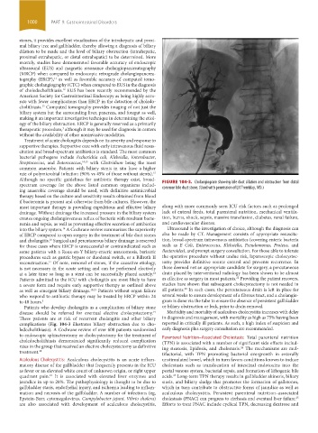
Although no specific guidelines for antibiotic therapy exist, broad- FIGURE 104-3. Cholangiogram showing bile duct dilation and obstruction from distal
spectrum coverage for the above listed common organisms includ-
ing anaerobic coverage should be used, with definitive antimicrobial common bile duct stone. (Used with permission of LN Tremblay, MD.)
therapy based on the culture and sensitivity results obtained from blood
if bacteremia is present and otherwise from bile cultures. However, the
most important therapy is providing expeditious and effective biliary along with more commonly seen ICU risk factors such as prolonged
drainage. Without drainage the increased pressure in the biliary system lack of enteral feeds, total parenteral nutrition, mechanical ventila-
creates ongoing cholangiovenous reflux of bacteria with resultant bacte- tion, burns, shock, sepsis, massive transfusion, diabetes, renal failure,
remia and sepsis, as well as preventing effective secretion of antibiotics and cardiovascular disease.
into the biliary system. A Cochrane review summarizes the superiority Ultrasound is the investigation of choice, although the diagnosis can
14
of ERCP compared to open surgery in the treatment of bile duct stones also be made by CT. Management consists of appropriate resuscita-
and cholangitis. Surgical and percutaneous biliary drainage is reserved tion, broad-spectrum intravenous antibiotics (covering enteric bacteria
16
for those cases where ERCP is unsuccessful or contraindicated such as such as E Coli, Enterococcus, Klebsiella, Pseudomonas, Proteus, and
some patients with a Roux-en-Y biliary-enteric anastomosis, bariatric Bacteroides), and prompt surgery consultation. For those able to tolerate
procedures such as gastric bypass or duodenal switch, or a Billroth II the operative procedure without undue risk, laparoscopic cholecystec-
reconstruction. Of note, removal of stones, if the causative etiology, tomy provides definitive source control and prevents recurrence. In
17
is not necessary in the acute setting and can be performed electively those deemed not an appropriate candidate for surgery, a percutaneous
at a later time so long as a stent can be successfully placed acutely. drain placed by interventional radiology has been shown to be almost
18
22
Patients admitted to the ICU with cholangitis are most likely to have as effective as surgery in most patients. Providing the patient recovers,
a severe form and require early supportive therapy as outlined above studies have shown that subsequent cholecystectomy is not needed in
23
as well as emergent biliary drainage. 10,19 Patients without organ failure all patients. In such cases, the percutaneous drain is left in place for
who respond to antibiotic therapy may be treated by ERCP within 24 several weeks to ensure development of a fibrous tract, and a cholangio-
to 48 hours. 5 gram is done via the tube to ensure the absence of persistent gallbladder
Patients who develop cholangitis as a complication of biliary stone or biliary obstruction or leak, prior to drain removal.
disease should be referred for eventual elective cholecystectomy. Morbidity and mortality of acalculous cholecystitis increases with delay
19
These patients are at risk of recurrent cholangitis and other biliary in diagnosis and management, with mortality as high as 75% having been
complications (Fig. 104-3 illustrates biliary obstruction due to cho- reported in critically ill patients. As such, a high index of suspicion and
ledocholithiasis). A Cochrane review of over 600 patients randomized early diagnosis plus surgery consultation are recommended.
to endoscopic sphincterotomy or cholecystectomy for the treatment of Parenteral Nutrition–Associated Cholestasis: Total parenteral nutrition
choledocholithiasis demonstrated significantly reduced complication (TPN) is associated with a number of significant side effects includ-
rates in the group that received an elective cholecystectomy as definitive ing steatosis, lipidosis, and cholestasis. The mechanisms are mul-
24
treatment. 20 tifactorial, with TPN promoting bacterial overgrowth in enterally
Acalculous Cholecystitis: Acalculous cholecystitis is an acute inflam- unstimulated bowel, which in turn favors conditions known to induce
matory disease of the gallbladder that frequently presents in the ICU cholestasis such as translocation of intestinal endotoxins into the
as fever or an elevated white count of unknown origin, or right upper portal venous system, bacterial sepsis, and formation of lithogenic bile
quadrant pain. It is associated with elevated liver enzymes and acids. Long-term TPN therapy results in gallbladder akinesis, biliary
25
21
jaundice in up to 20%. The pathophysiology is thought to be due to stasis, and biliary sludge that promotes the formation of gallstones,
gallbladder stasis, endothelial injury, and ischemia leading to inflam- which in turn contribute to obstructive forms of jaundice as well as
mation and necrosis of the gallbladder. A number of infections (eg, acalculous cholecystitis. Persistent parenteral nutrition– associated
Epstein-Barr, cytomegalovirus, Campylobacter jejuni, Vibrio cholera) cholestasis (PNAC) can progress to cirrhosis and eventual liver failure.
25
are also associated with development of acalculous cholecystitis, Efforts to treat PNAC include cyclical TPN, decreasing dextrose and
section09.indd 1000 1/14/2015 9:27:05 AM