
Page 1612 - Hall et al (2015) Principles of Critical Care-McGraw-Hill
P. 1612
CHAPTER 118: Head Injury 1131
■
The methods devised to quantitatively measure CBF were developed )
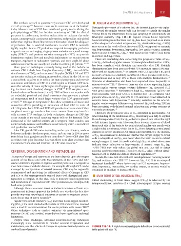
JUGULAR VENOUS BULB OXIMETRY (Sj O2
over 65 years ago ; however, none are in common use at the bedside Retrograde placement of a catheter into the internal jugular vein cepha-
120
today. Measurement of CBF has contributed greatly to elucidating the lad toward the jugular venous bulb can be used to sample the jugular
pathophysiology of TBI, but bedside monitoring of CBF for clinical venous blood via intermittent blood gas sampling or continuously via
purposes is cumbersome, involves radioactivity or indicator dye tech- fiberoptic oximetry (Fig. 118-14). Jugular venous blood represents
niques, cannot provide continuous information, and provides only global has been proposed as an index
rather than regional blood flow without information about the adequacy blood returning from the brain and Sj O 2 desaturations (eg, <55%)
of perfusion, that is, cerebral metabolism, to which CBF is normally of the adequacy of cerebral oxygenation. Sj O 2
may occur as the result of local (increased ICP, vasospasm) or systemic
tightly coupled. Xenon-CT, perfusion-computed tomography, perfusion (eg, hypotension, hypoxemia, hypocarbia, low cardiac output, anemia)
magnetic resonance imaging, single-photon emission-computed tomog- (eg, >74%) occur as the result of local tissue
raphy (SPECT), and positron emission tomography (PET) can provide factors as can elevated Sj O 2
infarction and hyperemia.
intermittent regional flow and metabolic information but require patient .
transport, expensive or radioactive materials, and even single, let alone There are conflicting data concerning the prognostic value of Sj O 2
, defined as jugular venous oxyhemoglobin desaturation <50%,
serial measurements, are usually not feasible in critically ill patients. 122,123 Low Sj O 2
has been correlated with disability and mortality after TBI. Multiple
More practical techniques currently being used at the bedside to desaturations have been associated with 71% mortality versus 18% mor-
estimate CBF include laser Doppler flowmetry (LDF), thermal diffu- tality with no desaturations. In patients with GCS ≤8 after TBI, good
127
sion flowmetry (TDF), and transcranial Doppler (TCD). LDF and TDF
are invasive techniques utilizing microprobes, placed in the OR or via recovery or moderate disability occurred in 44% of patients with no Sj O 2
128
desaturations and in only 15% of those with multiple desaturations.
a cranial bolt, adjacent to or within the brain parenchyma and provide Episodes of desaturation also have been reported more frequently in
continuous estimations of CBF in a small region of interest. LDF mea- nonsurvivors of TBI. However, there are also studies reporting higher
129
sures the velocity of tissue erythrocytes via Doppler principles provid- )
ing fractional (not absolute) changes in CBF. LDF samples a very arterio-jugular venous oxygen content difference (eg, decreased Sj O 2
124
saturation (≥75%) has
130
limited volume of brain tissue (1 mm ). TDF utilizes a microprobe with with good outcomes. Furthermore, high Sj O 2
3
been associated with poor GOS at 6 months post-TBI compared with
an embedded proximal and distal thermistor that generates a spheri- was 56% to 74% and may reflect infarcted or
cal temperature field of much larger volume than LDF, approximately patients whose mean Sj O 2 131
necrotic tissue with hyperemia. Similarly, reduction in the arterial-
27 mm . Changes in temperature flux allow separation of tissue and ) following TBI has
3 125
convective effects providing an estimation of local CBF in units of jugular venous oxygen difference (eg, increased Sj O 2
been associated with delayed cerebral infarction and poorer outcome at
mL/100 g/min. Both LDF and TDF will provide inaccurate data if they 6 months postinjury. 132
are placed over large vessels or lose tissue contact. Fever may interfere saturation is questionable. An
with accurate TDF readings. In both techniques, changes in CBF that Therefore, the prognostic value of Sj O 2
monitoring can help to explain
occur outside of the small sampling region will not be detected. TCD understanding of the limitations of Sj O 2
catheter is placed into either the right
ultrasound of the cerebral arteries via a temporal bone window can these discrepancies. First, the Sj O 2
or left internal jugular vein. However, there is some crossover of blood
noninvasively measure cerebral blood vessel velocity which is an indirect from each side of the brain to the contralateral jugular vein usually with
and qualitative index of CBF. from detecting contralateral
After TBI, global CBF varies depending on the type of injury, tends to a right-sided dominance, which limits Sj O 2
changes in oxygen saturation. Of paramount importance is the inability
be lowest in the first few hours posttrauma, and can vary by 25% or more measurement to determine whether the oxygen supply to brain
from lobar, basal ganglion and brain stem flow. Lower CBF after TBI of Sj O 2 can occur when the brain is able to extract
126
correlates with poor outcome; however, there is no clear evidence that cells is adequate. A low Sj O 2 , as noted above, may
measurement and directed treatment of CBF alter outcome. 85 enough oxygen to meet its needs and a high Sj O 2
indicate tissue infarction or hyperoxemia. A normal range Sj O 2 (eg,
<55%-74%) may only reflect the global mix and thus fail to detect
CEREBRAL OXYGENATION AND METABOLISM regional cerebral compromise. Therefore, the Sj O 2 value, without simul-
taneous CBF or metabolic data, is of limited significance. 84
Transport of oxygen and nutrients to the brain depends upon the oxygen To date, there is a lack of level I or II investigations of restoring normal
content of the blood and CBF. Measurements of ICP, CPP, and CBF Sj O 2 and outcome after TBI. 132,133 However, Sj O 2 <50 % is an accepted
cannot determine if global or regional cerebral perfusion is adequate to treatment threshold where factors such as ICP, vasospasm, hypoten-
84
meet brain tissue metabolic demand. For example, although CPP can be sion, hypoxemia, hypocarbia, low cardiac output, or anemia would be
managed by manipulation of arterial pressure, CBF may be regionally optimized in an effort to increase the Sj O 2 .
compromised and predicting the differential effects of changes in CBF
and ICP in the heterogeneously injured brain with dysregulated auto- ■ BRAIN TISSUE OXYGEN MONITORING (Pbt O2 )
regulation is complex. In this area, tools to measure tissue oxygenation ) is achieved by the
and metabolism in conjunction with other parameters, for example, ICP, Direct monitoring of brain tissue oxygen (Pbt O 2
intraparenchymal insertion of a Clark polarographic oxygen sensing
hold some promise.
Although there are several direct or indirect monitors of brain oxy-
genation and ischemia approved for bedside use, whether the data they
provide improves neurological outcomes or assists in prognosis contin-
ues to be an area of active research. 84
) and brain tissue oxygen monitor-
Jugular venous bulb oximetry (Sj O 2
), the most studied as they relate to TBI and outcome, received
ing (Pbt O 2
only a level III recommendation for use in patients with severe TBI.
84
Other techniques such as cerebral oximetry via near-infrared spec-
troscopy (NIRS) and cerebral microdialysis have significant technical
limitations.
Despite these challenges, advanced neuromonitoring techniques
increasingly allow intensivists to monitor cerebral oxygenation and
metabolism, and the effects of changes in systemic hemodynamics on FIGURE 118-14. A single lumen retrograde jugular bulb catheter (arrow) was inserted
cerebral hemodynamics. in this patient with acute TBI.
section10.indd 1131 1/20/2015 9:20:20 AM