
Page 1607 - Hall et al (2015) Principles of Critical Care-McGraw-Hill
P. 1607
1126 PART 10: The Surgical Patient
Mechanical ventilation causes positive intrathoracic pressure and higher are no studies performed to date that support a particular target blood
pressures can cause decreased venous return and a rise in jugular venous pressure. An analysis of the relationship between admission SBP and
pressure leading to an increase in cerebral blood volume (CBV) and in MAP after TBI and GOS (Fig. 118-10) at 6 months using the IMPACT
33
ICP and to a drop in cardiac output and blood pressure, thereby reducing database found that SBP on the order of 135 mm Hg and MAP on the
cerebral perfusion pressure (CPP) and cerebral blood flow (CBF). In areas order of 90 mm Hg were associated with the best outcome, although
34
where cerebral autoregulation is intact, decreases in CPP are compensated these data do not support a strong causal inference. However, because of
for by cerebral vasodilation, increasing CBV and potentially increasing ethical considerations, there are no class I studies (ie, well-designed ran-
ICP; if autoregulation is impaired, decreased CPP may lead to cerebral domized controlled trials) of the effect of blood pressure resuscitation
ischemia. The effect of these changes on the brain is difficult to impossible targets on outcome. As such, level II evidence (Fig. 118-11) supports
35
15
to monitor, but avoiding extremes and maintaining homeostasis is critical. a threshold systolic blood pressure of 90 mm Hg. A SBP of less than
15
In the TBI patient, premature extubation may result in 2nd injury. 26 90 mm Hg must be avoided if possible, or rapidly corrected.
Tracheostomy either by open or percutaneous dilational techniques, The type of hemodynamic monitoring employed should be determined
depending on patient anatomy and local expertise, should be performed by the severity of TBI, the degree of instability, the response or lack of
in patients expected to require mechanical ventilation for greater than response to resuscitation, and the expertise of the critical care physician.
10 to 14 days. The exact timing of tracheostomy remains a matter of Foremost, no matter what type of monitoring is employed it must be
debate. Tracheostomy may decrease the number of ventilator days, but coupled with the clinical context including physical examination, intake
there is no evidence that it decreases ICU length of stay or pneumonia and output, pertinent labs including hemoglobin, renal function, lactate,
rates. 27,28 The benefits of tracheostomy include better oral care, improved ABG, CXR, ECG, CT (intracranial pathology), ICP, and the results of any
patient comfort, decreased self-extubation risk, allowance for less sedation, additional neuromonitors, for example, brain tissue oxygen, CBF, etc.
better communication (speaking valve), more aggressive weaning attempts, Hemodynamically stable patients may be monitored simply by con-
decreased dead space ventilation, and possibly a lower work of breathing. tinuous blood pressure via arterial catheter and ECG. In patients with
severe or persistent hypotension, shock, multiple organ dysfunction, and
HEMODYNAMIC MONITORING AND MANAGEMENT intracranial hypertension, the titration of fluid and vasoactive agents is
more challenging. Vasopressors in the setting of intravascular volume
Both hypotension and raised ICP are the leading causes of death in depletion may worsen cerebral ischemia and other organ perfusion and
severe TBI and are related to the severity of the brain injury as well as the excess fluid resuscitation may lead to worsening pulmonary edema,
systemic complications. Hypotension exclusively from TBI is a terminal hypoxemia, cerebral ischemia, and cerebral edema.
event due to herniation. Central venous pressure (CVP) traditionally has been used to assess
Both mortality rate and outcome (ie, degree of disability) are signifi- the adequacy of intravascular volume and although it is still often mea-
cantly increased in patients with documented episodes of hypoxemia sured and discussed in neurosurgical ICU settings, it should not be used
16
or hypotension. An analysis of the large, prospectively collected, to guide fluid management. Recent studies have failed to demonstrate a
29
36
observational data set, the Traumatic Coma Data Bank (TCDB), found clinically useful correlation between absolute CVP or change in CVP with
that hypoxia and hypotension were independently associated with sig- intravascular volume or right ventricular preload, and it does not predict
37
nificant increases in morbidity and mortality in the setting of severe fluid responsiveness. The pulmonary artery occlusion pressure obtained
36
head injury. A single prehospital episode of systolic blood pressure from the pulmonary artery catheter (PAC) also does not provide accurate
30
<90 mm Hg is associated with increased morbidity and a doubling of information about left ventricular preload or intravascular volume. 37
mortality compared with a matched group of patients without hypo- Recent trends in hemodynamic monitoring in the critically ill favor
tension. Hypotension is among the five most powerful predictors of the use of less invasive and more direct measures of cardiac function 38,39
30
outcome after TBI, independent of the other major predictors of out- and dynamic indices predictive of preload (fluid) responsiveness.
40
come including age, admission GCS score, admission GCS motor score, Bedside echocardiography can rapidly and directly assess both right and
intracranial diagnosis, and pupillary status. In the hospital, repeated left ventricular preload and contractility and in trauma patients can rule
30
episodes of hypotension and increased total duration of hypotensive out significant pericardial effusion. The lungs can be assessed by ultra-
39
episodes were significant predictors of both mortality and poor neuro- sound on the same examination, to detect pulmonary edema (B-lines)
logical outcome. Patients that respond to resuscitation after TBI with early on, rapidly rule out a pneumothorax (presence of lung sliding) or
31
improved BP have a better survival. 32 hemothorax, and can detect atelectasis (shift of heart, echogenic lung
Hemodynamic management should employ fluids, vasoactive agents, appearance). Ultrasound can also be used to determine the inferior
39
and blood transfusions as indicated to maintain a systolic blood pres- vena cava (IVC) diameter and variability with respiration as a dynamic
sure above 90 mm Hg. The 90 mm Hg systolic pressure threshold is index of fluid responsiveness, although it may be inaccurate in the
40
derived from statistical distributions of blood pressure for normal setting of intra-abdominal hypertension, RV dysfunction, or pericardial
adults. Systolic blood pressures lack a consistent relationship with mean tamponade. Pulse contour analysis of the arterial pressure waveform can
arterial pressure (MAP) and MAP is used to calculate the cerebral perfu- measure the pulse pressure variability with respiration. Values greater
sion pressure (CPP). It may be desirable to maintain MAP considerably than 12% to 13% are more predictive of fluid responsiveness ; how-
41
above those represented by systolic pressures of 90 mm Hg, but there ever, the cardiac rhythm must be sinus, the tidal volumes constant and
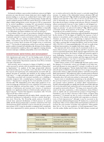
Score Rating Explanation
5 Good recovery Can resume normal life, minor deficits
4 Moderate disability Independent, e.g., travel by public
transport; work in sheltered setting
3 Severe disability Dependent for daily support
2 Persistent vegetative state Partial arousal, but lack any awareness
1 Dead
FIGURE 118-10. Glasgow Outcome Scale (GOS). (Data from Jennett B, Bond M. Assessment of outcome after severe brain damage, Lancet. March 1, 1975;1(7905):480-484.)
section10.indd 1126 1/20/2015 9:20:16 AM