
Page 1608 - Hall et al (2015) Principles of Critical Care-McGraw-Hill
P. 1608
CHAPTER 118: Head Injury 1127
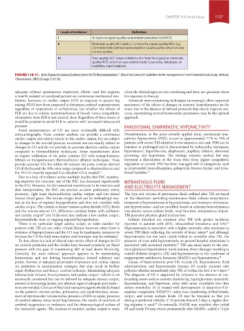
Levels of evidence Definition
I At least one good quality randomized controlled trial (RCT).
II Moderate quality RCT; lacks ≥1 criteria for a good quality RCT, e.g.,
controlled trials without randomization. Good quality cohort or case-
control studies.
III Poor quality RCT; major violations of criteria for a good or moderate
quality RCT, cohort or case control study; Case series, databases, or
registries, expert opinion
35
FIGURE 118-11. Brain Trauma Foundation Evidence Levels for TBI Recommendations. (Data from Carney NA. Guidelines for the management of severe traumatic brain injury. Methods,
J Neurotrauma. 2007;(24 suppl 1):S3-S6).
adequate without spontaneous respiratory efforts—and this requires when the clinical signs are not correlating and there are questions about
a heavily sedated or paralyzed patient on continuous mechanical ven- the response to therapy.
tilation. Increases in cardiac output (CO) in response to passive leg Advanced neuromonitoring techniques increasingly allow improved
raising (PLR) have been proposed to determine preload responsiveness assessment of the effects of changes in systemic hemodynamics on the
regardless of respirations or arrhythmias, but whether the effects of brain, but in the absence of defined protocols that clearly improve out-
PLR are due to volume (autotransfusion of blood) versus sympathetic come, maintaining normal homeostatic parameters may be the optimal
stimulation from PLR is not entirely clear. Regardless of these issues, it approach.
would be prudent to avoid PLR in patients with increased intracranial
pressure. PAROXYSMAL SYMPATHETIC HYPERACTIVITY
Serial measurements of CO are more technically difficult with
echocardiography. Pulse contour analysis can provide a continuous Dysautonomia, or the more recently applied term, paroxysmal sym-
cardiac output and relative trends in the cardiac output, but are subject pathetic hyperactivity (PSH), occurs in approximately 7.7% to 33% of
to changes in the arterial pressure waveform not necessarily related to patients with severe TBI admitted to the intensive care unit. PSH can be
changes in CO and do not provide an accurate absolute cardiac output transient or prolonged and is characterized by tachycardia, tachypnea,
compared to thermodilution techniques. Some manufactures allow hypertension, hyperthermia, diaphoresis, pupillary dilation, abnormal
or require calibration of the pulse contour CO with transpulmonary posturing, and hypertonia. The etiology remains unclear, but may
lithium or transpulmonary thermodilution dilution techniques. These represent a dissociation of the brain stem from higher sympathetic
provide accurate CO, but within 60 minutes the pulse contour-derived regulation or control. PSH has been managed with β-antagonists, such
CO drifts beyond the 30% error range compared to thermodilution and as propranolol, benzodiazepines, gabapentin, bromocriptine, and intra-
the TD CO must be repeated if an absolute CO is needed. 42 thecal baclofen. 44
Due to a lack of evidence across multiple studies that PAC monitor-
ing improves the outcome, use of the PAC has decreased significantly INTRAVENOUS FLUID
in the ICU. However, for the intensivist experienced in its insertion and AND ELECTROLYTE MANAGEMENT
data interpretation, the PAC can provide accurate pulmonary artery
pressures, right heart thermodilution cardiac output, and true mixed The type and volume of intravenous fluids utilized after TBI are based
venous blood gases. The venous oxygen level can be misleadingly nor- on the objectives—providing maintenance fluid, volume resuscitation,
mal in the face of regional hypoperfusion and does not correlate with treatment of hypernatremia or hyponatremia, and treatment of intracra-
cardiac output. The central or mixed venous carbon dioxide (CO ) levels nial hypertension—and are modified based on systemic hemodynamics
2
and the venous-arterial CO difference correlate better with perfusion (see above), serum sodium levels, renal function, and presence of post-
2
and cardiac output and if elevated may indicate a low cardiac output, TBI posterior pituitary gland dysfunction.
43
hypermetabolic state, or ongoing regional hypoperfusion. Sodium disorders are common after TBI with greater incidence
There is no particular target cardiac output or index number for reported in patients with SDH, intracerebral hematoma and DAI.
45
patients with TBI (or any other critical illness); however, when there is Hypernatremia is associated with a higher mortality after moderate to
46
evidence of hypoperfusion and the CO may be inadequate, measures to severe TBI likely reflecting the severity of brain injury and although
increase the CO by fluid resuscitation and inotropes may be instituted. hyponatremia has not been clearly linked to mortality after TBI, the
To date, there is a lack of clinical data on the effect of changes in CO presence of even mild hyponatremia on general hospital admissions is
on cerebral perfusion and the studies have focused primarily on blood associated with increased mortality. TBI can cause injury to the pitu-
47
pressure with the goal of maintaining SBP at least above 90 mm Hg. itary gland and hypothalamic tracts (edema, direct damage) resulting in
Currently the most rational approach appears to be maintaining central diabetes insipidus (DI) and hypernatremia or the syndrome of
homeostasis and not driving hemodynamics toward arbitrary end inappropriate antidiuretic hormone (SIADH) and hyponatremia. 48
points. Normal or adequate parameters of pressure and cardiac output Causes of hypernatremia post-TBI include DI, hypernatremic fluid
are preferable to maximization strategies that may result in further administration, and hyperosmolar therapy. DI usually presents with
organ dysfunction and hence, cerebral ischemia. Maintaining adequate polyuria whether immediately after TBI or within the first 2 to 3 days.
49
intravascular volume, blood pressure, and cardiac output—which is not The diagnosis of DI is supported by polyuria in the absence of con-
necessarily monitored but can be inferred by adequate urinary output, founding causes such as osmotic diuresis (eg, hyperglycemia, mannitol),
normal or decreasing lactate, and physical signs of adequate perfusion— hypernatremia, and hypotonic urine with urine osmolality less than
is recommended. Choices of fluid and vasoactive agents should be based serum osmolality. DI is treated with desmopressin (1-desamino-8-D-
on the patient’s current cardiac, pulmonary, and renal function, assess- arginine vasopressin [DDAVP]) under close monitoring of fluid intake,
ment of intravascular volume status, presence of SIRS or sepsis, presence output, and serum sodium levels. DI may be transient so that prn
of cerebral edema, intracranial hypertension, the results of monitors of dosing is preferred initially; if DI persists beyond 2 days a regular dos-
cerebral oxygenation or perfusion, and the pharmacological actions of ing regimen is used. Occasionally, SIADH may manifest after initial
49
the vasoactive agents. The decision to monitor cardiac output is made DI and rarely DI may return permanently after SIADH—called a “triple
section10.indd 1127 1/20/2015 9:20:17 AM