
Page 1628 - Hall et al (2015) Principles of Critical Care-McGraw-Hill
P. 1628
CHAPTER 119: Spinal Injuries 1147
1 2 3
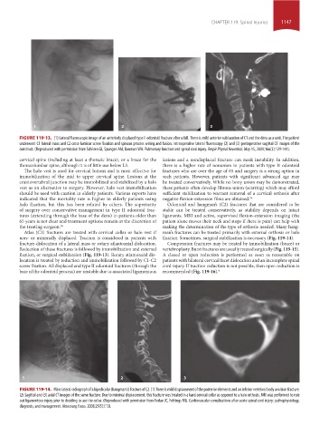
FIGURE 119-13. (1) Lateral fluoroscopic image of an anteriorly displaced type II odontoid fracture after a fall. There is mild anterior subluxation of C1 and the dens as a unit. The patient
underwent C1 lateral mass and C2 cross-laminar screw fixation and spinous process wiring and fusion. Intraoperative lateral fluoroscopy (2) and (3) postoperative sagittal CT images of the
construct. (Reproduced with permission from Schilero GJ, Spungen AM, Bauman WA. Pulmonary function and spinal cord injury. Respir Physiol Neurobiol. May 15, 2009;166(3):129-141).
cervical spine (including at least a thoracic brace), or a brace for the lesions and a nondisplaced fracture can mask instability. In addition,
thoracolumbar spine, although it is of little use below L3. there is a higher rate of nonunion in patients with type II odontoid
The halo vest is used for cervical lesions and is most effective for fractures who are over the age of 65 and surgery is a strong option in
immobilization of the mid to upper cervical spine. Lesions at the such patients. However, patients with significant advanced age may
craniovertebral junction may be immobilized and stabilized by a halo be treated conservatively. While no bony union may be demonstrated,
vest as an alternative to surgery. However, halo vest immobilization these patients often develop fibrous union (scarring) which may afford
should be used with caution in elderly patients. Various reports have sufficient stabilization to warrant removal of a cervical orthosis after
indicated that the mortality rate is higher in elderly patients using negative flexion-extension films are obtained. 56
halo fixation, but this has been refuted by others. The superiority Odontoid and hangman’s (C2) fractures that are considered to be
of surgery over conservative management in type II odontoid frac- stable can be treated conservatively, as stability depends on intact
tures (extending through the base of the dens) in patients older than ligaments. MRI and active, supervised flexion-extension imaging (the
65 years is not clear and treatment options remain at the discretion of patient alone moves their neck and stops if there is pain) can help with
the treating surgeon. 56 making the determination of the type of orthosis needed. Many hang-
Atlas (C1) fractures are treated with cervical collar or halo vest if man’s fractures can be treated primarily with external orthosis or halo
non- or minimally displaced. Traction is considered in patients with fixation. Sometimes, surgical stabilization is necessary (Fig. 119-14).
fracture-dislocation of a lateral mass or rotary atlantoaxial dislocation. Compression fractures may be treated by immobilization (brace) or
Reduction of these fractures is followed by immobilization and external vertebroplasty. Burst fractures are usually treated surgically (Fig. 119-15).
fixation, or surgical stabilization (Fig. 119-13). Rotary atlantoaxial dis- A closed or open reduction is performed as soon as reasonable on
location is treated by reduction and immobilization followed by C1-C2 patients with bilateral cervical facet dislocation and an incomplete spinal
screw fixation. All displaced and type II odontoid fractures (through the cord injury. If traction reduction is not possible, then open reduction is
base of the odontoid process) are unstable due to associated ligamentous recommended (Fig. 119-16). 4
1 2 3
FIGURE 119-14. Plain lateral radiograph of a bipedicular (hangman’s) fracture of C2. (1) There is mild displacement of the posterior elements and an inferior vertebral body avulsion fracture.
(2) Sagittal and (3) axial CT images of the same fracture. Due to minimal displacement, this fracture was treated in a hard cervical collar as opposed to a halo orthosis. MRI was performed to rule
out ligamentous injury prior to deciding to use the collar. (Reproduced with permission from Furlan JC, Fehlings MG. Cardiovascular complications after acute spinal cord injury: pathophysiology,
diagnosis, and management. Neurosurg Focus. 2008;25(5):E13).
section10.indd 1147 1/20/2015 9:20:40 AM