
Page 1623 - Hall et al (2015) Principles of Critical Care-McGraw-Hill
P. 1623
1142 PART 10: The Surgical Patient
A
B
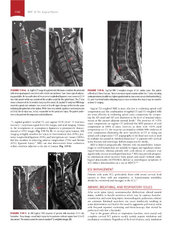
FIGURE 119-6. A. Sagittal CT image of a patient who fell down a manhole. He presented FIGURE 119-8. Sagittal MRI T2-weighted images of the lumbar spine. This patient
with dense quadraparesis consistent with central cord syndrome. Scan shows signs of ankylos- suffered an L1 burst fracture. There is increased signal intensity within the L1 body indicating
ing spondylitis. He has ossification of the posterior longitudinal ligament, most severe at C3-C4 contusion/edema. In addition, higher signal intensities (see arrow) can also be found in the L2,
(see thick arrow) which was considered the possible culprit for the spinal injury. The CT also L3, and L4 vertebral bodies indicating injury to those vertebrae that may or may not manifest
reveals a fracture of the C6 vertebral body (see thin arrow). B. Sagittal T2-weighted MRI image on bone CT imaging.
revealed a spinal cord contusion (see arrow) at C6 with signal changes within the disc space
indicating disruption but no herniation. While there was, indeed, significant cord compression Sagittal T2-weighted MRI is most effective at evaluating spinal cord
at C3-C4, the C6 injury was, in fact, responsible for the spinal cord injury. The patient under- compression and the combination of sagittal CT and T1-weighted MRI
went early posterior decompression and stabilization. are most effective at evaluating spinal canal compromise by compar-
ing the AP canal and AP cord diameters at the level of maximal injury
versus at the nearest adjacent normal levels. The presence of >25%
4
T1, sagittal gradient recalled T2, and sagittal STIR (short TI inversion
recovery—suppresses signal from fat) images, and axial imaging. Edema canal compromise on sagittal CT predicted the MRI presence of cord
compression in 100% of cases; however, in those with <25% canal
in the interspinous or supraspinous ligaments is prominently demon-
strated on STIR images (Fig. 119-7A, B). In cervical spine trauma, MR compromise on CT, the majority continued to exhibit MRI evidence of
cord compression illustrating the poor specificity of CT in ruling out
imaging is highly sensitive for injury to intervertebral disk (93%), pos- 4
terior longitudinal ligament (93%), and interspinous soft tissues (100%), spinal cord compression. CT angiography of the head and neck is used
to evaluate for carotid or vertebral dissection in patients with cervical
18
but less sensitive in detecting anterior longitudinal (71%) and flavum spine fracture and neurologic deficits suspicious for stroke.
(67%) ligament injury. MRI can also demonstrate bone contusions MRI is helpful prognostically. Patients with intramedullary hemor-
17
within vertebrae adjacent to the site of trauma (Fig. 119-8).
rhage or cord transsection are unlikely to regain any significant neuro-
logical function, whereas patients with cord edema or contusion may
significantly recover neurological function. MRI may provide prognos-
19
tic information about recovery from spinal cord injury without radio-
logical abnormality (SCIWORA) defined as neurological symptoms of
SCI without abnormalities on x-ray or MDCT. 4,20
A
ICU MANAGEMENT
Patients with acute SCI, particularly those with severe cervical level
injuries or those with any respiratory or hemodynamic instability,
should be monitored and managed in the ICU. 14
AIRWAY, BREATHING, AND RESPIRATORY ISSUES
After acute spine injury, neuromuscular dysfunction, altered mental
status, inability to handle secretions which may be copious, broncho-
constriction, and acute lung injury (noncardiogenic pulmonary edema)
are common. Retained secretions can occur insidiously resulting in
acute deterioration and lead to the need for aggressive pulmonary toilet
with frequent repeated suctioning and bronchoscopy that should be
performed with low a threshold.
FIGURE 119-7. A. MR Sagittal STIR sequence of patient with traumatic C3-C4 disc Due to the greater effects on respiratory function, more rostral and
herniation. These images reveal bright signal in the posterior subfacial region from C3 to C5 complete cervical SCI patients usually acutely require intubation and
(see arrow). This edema cannot be seen on standard T2-weighted images (B). mechanical ventilation. Regardless of the level of injury, early intubation
section10.indd 1142 1/20/2015 9:20:30 AM