
Page 1624 - Hall et al (2015) Principles of Critical Care-McGraw-Hill
P. 1624
CHAPTER 119: Spinal Injuries 1143
under controlled circumstances is preferred to avoid secondary injuries noncardiogenic pulmonary edema (acute respiratory distress syndrome
resulting from hypoxemia or respiratory system-induced hemodynamic [ARDS]) leading to hypoxemic respiratory failure, and decreased sym-
failure. Evidence of respiratory failure includes altered mental status, pathetic tone (with increased relative parasympathetic tone) leading
hypoxemia, rapid, shallow, or irregular breathing with associated respi- to bronchoconstriction and increased airway secretions. Sympathetic
(late) or a progressive decline in (bronchodilatory) innervation of the lungs arises from the T1-T6 level so
ratory alkalosis (early) or elevated P CO 2
serial vital capacity (VC), or VC <1.0 L. that tetraplegic (ie, cervical) SCI leads to loss of sympathetic innervation
In critically ill patients in general, and trauma and SCI patients in to the lungs and unopposed or increased parasympathetic vagal activity
particular, tracheal intubation is significantly more difficult due to factors that can result in decreased baseline airway caliber. The use of anti-
27
such as the need for precautionary neck stabilization, bleeding, vomiting, cholinergic bronchodilators such as ipratropium may be considered ;
28
oropharyngeal secretions, respiratory dysfunction, airway edema, however, it is not known if the baseline bronchoconstriction in
hemodynamic instability, and encephalopathy. Halo traction devices are tetraplegia contributes to respiratory symptoms. Aggressive suctioning
27
not readily removable and also limit the ability to position the airway for of secretions can result in bronchial stimulation and increased para-
intubation, increasing difficulties. Bradycardia and hypotension during sympathetic output resulting in bradyarrhythmia or conduction blocks.
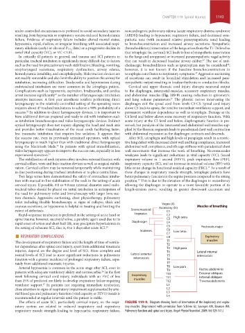
endotracheal intubation are more common in the tetraplegic patient. Cervical and upper thoracic cord injury disrupts neuronal output
Complications such as hypoxemia, aspiration, bradycardia, and cardiac to the diaphragm, intercostal muscles, accessory respiratory muscles,
arrest increase significantly as the number of laryngoscopic intubation and abdominal muscles (Fig. 119-9) causing reduction in spirometric
21
attempts increases. A first year anesthesia resident performing direct and lung volume parameters. The phrenic nerves innervating the
27
laryngoscopy in the relatively controlled setting of the operating room diaphragm exit the spinal cord from levels C3-C5. Spinal cord injury
requires about 47 tracheal intubations to achieve a 90% probability of a above C3 leads to apnea, the need for immediate ventilatory support, and
success. In addition to direct laryngoscopy devices, it is important to permanent ventilator dependence as well as tetraplegia. Damage at the
22
have additional devices prepared and ready to aid with intubation such C4 level and below allows some recovery of respiratory function. With
as intubation bronchoscopes and video laryngoscopic devices. Indirect acute injury at the C5 level and below, diaphragmatic function is pre-
optical laryngoscopy does not require aligning the head and neck axis served, but paralysis of the intercostal and abdominal wall muscles sup-
and provides better visualization of the vocal cords facilitating faster, plied by the thoracic segments leads to paradoxical chest wall contraction
less traumatic intubation that requires less sedation. It appears that with abdominal expansion as the diaphragm contracts and descends.
the success rate, even in previously untrained operators, with optical After SCI, tetraplegia and high levels of paraplegia result in a restric-
laryngoscopy is much higher than with traditional direct laryngoscopy tive lung defect with decreased chest wall and lung compliance, increased
using the Macintosh blade. In patients with spinal immobilization, abdominal wall compliance, and rib cage stiffness with paradoxical chest
23
video laryngoscopy appears to improve the success rate, especially in less wall movements that increase the work of breathing. Neuromuscular
experienced operators. 24 weakness leads to significant reductions in vital capacity (VC), forced
The stabilization of neck injuries often involves external fixation with expiratory volume in 1 second (FEV1), peak expiratory flow (PEF),
cervical collars, vests and halo traction devices as well as surgical stabili- inspiratory capacity (IC), and an increase in residual volume (RV) with
zation. Cervical collars may be removed temporarily while maintaining little or no change in functional residual capacity (FRC). As a result of
27
in-line positioning during tracheal intubation or to place central lines. these changes in respiratory muscle strength, tetraplegic patients have
Two large series have demonstrated the safety of orotracheal intuba- better pulmonary function in the supine position compared to the sitting
tion with manual in-line stabilization of the neck in the setting of acute position. This is due to the elevation of the diaphragm in recumbency
27
cervical injury. If possible, 8.0 or 9.0 mm internal diameter sized endo- allowing the diaphragm to operate in a more favorable portion of its
tracheal tubes should be placed on initial intubation in anticipation of length-tension curve, resulting in greater downward excursion and
the need for pulmonary toilet and bronchoscopy with larger bore suc-
tion channels. Aggressive suctioning, chest physiotherapy, pulmonary
toilet including flexible bronchoscopy at signs of collapse, thick and
Vagus (X)
copious secretions, or hypoxemia is helpful in treating and limiting the Accessory (XI) Muscles of breathing
extent of atelectasis. Sternomastoid C1
Rapid-sequence intubation is preferred in the setting of acute head or trapezius C2
spine trauma; however, succinylcholine, a paralytic agent used due to its C3 High tetra
rapid onset of action and short half-life, may precipitate hyperkalemia in Diaphragm C4
the setting of subacute SCI, that is, 4 to 5 days after acute SCI. 25 C5 Pectoralis major
C6
■ RESPIRATORY COMPLICATIONS Scalenes C7 Low tetra Expiratory
C8
The development of respiratory failure and the length of time of ventila- T1 muscles
tor dependence after spinal cord injury, apart from additional traumatic T2
injuries, depend on the degree and level of SCI. More complete and T3 High para Lateral internal
T4
rostral levels of SCI lead to more significant reductions in pulmonary Lateral external T5 intercostals
function with a greater incidence of prolonged respiratory failure, sepa- intercostals T6
rately from additional traumatic injuries. T7
Arterial hypoxemia is common in the acute stage after SCI, even in T8 Low para
patients with adequate ventilatory ability and normocarbia. In the first T9 Rectus abdominis
26
External obliques
week following cervical cord injury, individuals with an FVC of less Inspiratory T10 Internal obliques
T11
than 25% of predicted are likely to develop respiratory failure requiring muscles T12 Traverseabdominis
ventilator support. In patients not requiring immediate intubation, L1
27
close attention to signs of respiratory impairment supplemented by arte- L2
rial blood gas and pulmonary function (vital capacity or FEV1) trends is L3
recommended at regular intervals until the patient is stable. L4
The effects of acute SCI, particularly cervical injury, on the respi- FIGURE 119-9. Diagram showing levels of innervation of the inspiratory and expira-
ratory system are related to reduced inspiratory and expiratory tory muscles. (Reproduced with permission from Schilero GJ, Spungen AM, Bauman WA.
respiratory muscle strength leading to hypercarbic respiratory failure, Pulmonary function and spinal cord injury. Respir Physiol Neurobiol. 2009;166:129-141.)
section10.indd 1143 1/20/2015 9:20:31 AM