
Page 1630 - Hall et al (2015) Principles of Critical Care-McGraw-Hill
P. 1630
CHAPTER 119: Spinal Injuries 1149
1 2 3
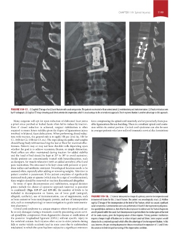
FIGURE 119-17. (1) Sagittal CT image of an L2 burst fracture with canal compromise. This patient was treated with an anterolateral L2 vertebrectomy and instrumentation. (2) Final construct as seen
by AP radiograph. (3) Sagittal CT image showing good direct anterior decompression, which is an advantage to the anterolateral approach. Short-segment fixation is another advantage to this approach.
Many surgeons will opt for open reduction of dislocated facet joints force compressing the spinal cord anteriorly and/or posteriorly from pos-
a priori since perched or locked facets often fail to reduce by traction. sible ligamentum flavum buckling. There is a resultant spinal cord contu-
Even if closed reduction is achieved, surgical stabilization is often sion within its central portion. Central cord syndrome can also be seen
required to assure future stability given the degree of ligamentous injury in younger patients who have suffered traumatic cervical disc herniations
involved in bilateral; facet dislocations. When performing closed reduc-
tion with traction, the general rule is to apply 5 lb per level (ie, 5 lb for
C1, 10 lb for C2, 15 lb for C3, etc). The rope along the pulley and weights
should hang freely without touching the bed or floor for maximal effec-
tiveness. Patients may or may not have shoulder rolls depending upon
whether the goal is to achieve extension, flexion, or simple distraction.
Hard collars are often maintained during traction for added stability
and the head of bed should be kept at 20° to 30° to avoid aspiration.
Awake patients are concomitantly treated with benzodiazepines, such
as diazepam, for muscle relaxation (with an added anxiolytic effect) and
pain medication. Pin sites need to be kept clean with peroxide or povi-
done iodine and antibiotic ointment. Neurological function needs to be
assessed often, especially after adding or removing weights. Attention to
patient comfort is paramount. If the patient complains of significantly
increased neck pain or spasms (which may be a sign of overdistraction),
then weights should be removed or traction discontinued altogether.
In terms of open decompression and stabilization, surgical decision
points include the choice of operative approach (anterior vs posterior
vs combined) (Figs. 119-17 and 119-18), the number of levels to be 1 2
included in decompression or fusion, use of bone graft (autograft,
allograft, synthetic), use of instrumentation, use of supplements, such FIGURE 119-18. (1) Lateral intraoperative image of a primary posterior decompression and
as bone cement or bone morphogenic protein, and use of intraoperative instrumented fusion for this L3 burst fracture. The patient was neurologically intact. (2) Midline
aids, such as neurophysiology or neuronavigation to guide instrumenta- sagittal CT image of the decompression at the level of the fracture, which has caused significant
tion placement. 4 canal compromise. Laminectomies were also performed at L4 and L5 due to preexisting degenera-
Central cord syndrome is a unique situation where the spinal cord can tive spondylolysis and stenosis. Note that the decompression is indirect and the fracture fragments
be acutely damaged during a traumatic event by preexisting severe cervi- are still present within the canal. Such decompression is permissible in the lumbar spine at the level
cal spondylitic compression from degenerative disease or ossification of of the cauda equina, given the forgiving nature of these injuries. Primary posterior stabilization
the posterior longitudinal ligament (OPLL) without specific injury to requires a longer length of fixation due to reduce delayed construct failure. Some surgeons would
the vertebral column. Such injuries often occur in older patients during choose to do a combined approach which offers the advantage of shorter segment fixation. In this
falls or motor vehicle accidents (and in some cases due to endotracheal case, however, the pre-existing degenerative disease necessitated incorporation of L5 and S1 into
intubation) in which the spine has been subject to a significant extension the construct to limit delayed worsening of the degenerative condition.
section10.indd 1149 1/20/2015 9:20:55 AM