
Page 1851 - Hall et al (2015) Principles of Critical Care-McGraw-Hill
P. 1851
1320 PART 11: Special Problems in Critical Care
diving is expensive but effective and relatively safe to depths of 400 fsw. preexisting medical conditions or injuries that compromise regional
Most dives deeper than 400 fsw require saturation of the diver with inert blood flow. Other serious forms of DCS involving the audiovestibular
gas at the approximate working depth of the dive. Saturation divers can system (staggers) and pulmonary system (chokes), although relatively
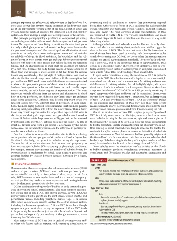
live and work for weeks at pressure, for instance in a bell and chamber rare, also occur. The most common clinical manifestations of DCS
system, and then undergo a single slow decompression to the surface. are presented in Table 132-2. The variable manifestations can make
The principle pathophysiologic problems of compressed gas diving the clinical diagnosis difficult to establish, and there are no diagnostic
occur during ascent due to the uncontrolled emergence of inert gas from laboratory studies.
tissues. During an ascent from diving or to altitude the extra inert gas in Although bubbles do lead to bends, some bubbles are clinically silent.
the body at the higher pressure is eliminated as the pressure decreases by As a result there is uncertainty about precisely how bubbles trigger the
the process of decompression. The rates of uptake or elimination of inert diverse features of DCS. The factors that govern bubble formation in
5
gases from the body after a pressure change are determined primarily by model tissues have been used to develop safe decompression tables
the solubility of the gas in blood and tissue, the blood flow, and the vol- usually by assuming that DCS will not occur unless the inert gas tension
ume of tissue. In most tissues, inert gas exchange follows an exponential exceeds the critical supersaturation threshold. The use of such a thresh-
function with respect to time. Tissues that behave this way are perfusion old is empirical, and in the subcritical range of supersaturation, DCS
limited, and the characteristics of their inert gas exchange are defined occurs as a stochastic event. Therefore, even appropriate use of well-
6
by a half-time. Because the body tissues receive different amounts of tested decompression tables or a decompression computer is associated
blood flow and nitrogen is more soluble in fat, half-times for various with a finite risk of DCS for dives deeper than about 25 fsw.
tissues vary considerably. The principle of multiple tissues was used to In open water recreational diving, the incidence of DCS is probably
calculate the first safe decompression tables, with the assumption that about one in 3000 dives, but increases with depth and duration, multiple
gas bubbles and DCS would occur only if the tissues were supersaturated same day dives, cold water and strenuous work. In military and commer-
to allow a nitrogen partial pressure of about twice the absolute pressure. cial divers and in military aviation, the risk is slightly higher, with a pre-
Modern decompression tables are still based on such parallel expo- dominance of mild to moderate type I symptoms. Tunnel workers have
nential models, but with lower degrees of supersaturation. The most a reported incidence of DCS of 0.7% to 1.5%, primarily consisting of
important variable affecting inert gas uptake and elimination is blood type I symptoms of the knee and lower leg. In recreational divers, some
7
flow or tissue perfusion, but diffusion may limit tissue gas exchange surveys have suggested frequent type II symptoms, but underreporting
under some conditions. Diffusion may become important when two of type I DCS and overdiagnosis of type II DCS are common. Delays
5
8
adjacent tissues have very different rates of perfusion. In such condi- in the diagnosis and treatment of DCS may also allow more severe
tions, the more highly perfused tissue eliminates inert gas more quickly, manifestations to evolve. Recreational divers are also more likely to omit
allowing inert gas to diffuse into it from the slower tissue. Thus, a faster decompression than are professional divers, thus increasing the risk.
tissue may remain supersaturated for longer than expected. Diffusion is One of the most serious forms of DCS is spinal paralysis. Spinal cord
9
also important during decompression once gas bubbles have formed in DCS is not fully understood, but the injury may be related to intravas-
a tissue. Bubbles contain large amounts of N 2 gas that can be removed cular bubbles forming in the low-pressure, epidural venous plexus of
by perfusion only after the N 2 diffuses back into the tissue. The rate at the spinal cord. Because of its low blood flow, the plexus is susceptible
10
which N 2 diffuses away from a gas bubble is determined by the bubble to bubble formation. Bubble-induced thrombi can obstruct venous out-
surface area, the intrabubble pressure, and the difference in partial pres- flow, leading to spinal cord ischemia. Despite evidence for bubble for-
sure between bubble and tissue. mation in the spinal venous plexus, intravascular formation of bubbles is
Bubbles tend to form in specific nucleation sites in the body during otherwise uncommon. Most intravascular bubbles probably originate at
decompression. Microscopic gas nuclei can be stabilized at hydropho- the tissue-blood interface and stream into the circulation to be absorbed
bic sites in the body, but grow into bubbles during decompression. by the lungs. Bubbles arising in the body of the spinal cord (autochtho-
The number of nucleation sites and their location and propensity to nous) have also been implicated in the etiology of spinal DCS. 11
form macroscopic bubbles differ according to physiologic conditions. Once bubbles enter the circulation, surface activity at the blood-
For example, exercise may increase the number of bubbles formed by to-bubble interface produces complement activation, activation of
tribonucleation, a mechanism by which large negative pressures can coagulation and fibrinolysis, platelet and neutrophil aggregation and
generate bubbles by traction between surfaces lubricated by a liquid,
such as joints.
■ DECOMPRESSION ILLNESS TABLE 132-2 Clinical Manifestations of DCS
Type I (mild DCS)
Decompression illness encompasses both decompression sickness (DCS) Limbs
and arterial gas embolism (AGE) and these conditions, particularly after Pain (bends), niggles, mild lymphatic obstruction, numbness, and paresthesias
an uncontrolled ascent by an inexperienced diver, may coexist. As a usually involving the large joints, eg, shoulders, elbows, and knees
rule, AGE has more serious implications, and it is a medical emergency. Skin
Arterial gas blocking cerebral or coronary vessels and causing ischemia Itching, rash, pallor, urticaria, edema (severe lymphatic obstruction, mottling,
must be eliminated promptly for the best outcome. and edema is considered serious [cutis marmorata])
DCS is attributable to the growth of bubbles in body tissues that pro-
duce one or more clinical manifestations. The most common presenta- Type II (serious DCS)
tion is pain-only or type I DCS, also known as bends. In type I DCS, the CNS
primary sites of bubble growth are the joint spaces, tendon sheaths, and Brain
periarticular tissues, including peripheral nerves. Type II or serious Headache, seizures, loss of consciousness, visual disturbances, hemiparesis,
DCS is less common and usually involves the central nervous system, aphasia, tremor, ataxia (staggers)
including the brain and spinal cord. Altitude DCS is similar, although Spinal cord
symptoms appear most often during the exposure. Altitude DCS tends Low back or pelvic girdle pain, paraparesis, urinary retention, incontinence
to be pain-only because the subject has often breathed an O 2-enriched Audiovestibular DCS
gas or has undergone O 2 prebreathing. Although uncommon, cases Tinnitus, vertigo, nystagmus, decreased hearing, nausea, and vomiting
involving the CNS do occur. Cardiopulmonary DCS (chokes)
Most serious cases of DCS are due to omitted decompression and/ Dyspnea, cough, wheezing, hemodynamic collapse
or other risk factors such as exercise, cold, coexisting dehydration, or CNS, central nervous system; DCS, decompression sickness.
section11.indd 1320 1/19/2015 10:56:10 AM