Page 625 - Clinical Hematology_ Theory _ Procedures ( PDFDrive )
P. 625

CHAPTER 29  ■  Body Fluid Analysis                        609


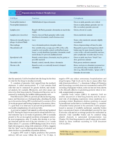
                       TABLE       29.18        Papanicolaou-Stained Morphology


                       Cell Type                                     Nucleus                                                                                  Cytoplasm


                       Neutrophils (mature)                          Multilobulated; hyperchromatic                                                           Green or pink; granules not evident


                       Neutrophils (immature)                                                                                                                 Green or pink; primary granules not vis-

                                                                                                                                                              ible; secondary granules present

                       Lymphocytes                                   Round with   nely granular chromatin; no nucleolus                                       Green; absent or scanty

                                                                     visible


                       Lymphocytes (reactive)                        Oval to cleaved;   nely granular with evenly                                             Green; moderate amount

                                                                       distributed chromatin; small chromocenter


                       Monocyte (usually macro-                      Lacy                                                                                     Some color; moderate amount; slightly

                       phages in pleural   uid)                                                                                                               vacuolated

                       Macrophage                                    Lacy chromatin pattern; irregular shape                                                  Green; degenerating cell may be pink.


                       Mesothelial cell                              Size variable (may occupy up to 50% of the cell);                                        Deep pink or green; homogeneous distri-

                                                                     round to oval; usually central; well-de  ned mem-                                        bution; may be more densely stained in

                                                                     brane; evenly distributed granular chromatin; small                                      center of the cell and around the nucleus;

                                                                     nucleoli may be present; multinucleated                                                  pale cytoplasmic vacuoles may be seen.


                       Ependymal cells                               Round; central dense chromatin; may be grainy or                                         Green or pink; may have “brush” bor-

                                                                     possible nucleoli                                                                        ders; generous amount


                       Choroidal cells                               Round; central, smooth, dense chromatin                                                  Pale green; moderate amount


                       Plasma cells                                  Round to oval; eccentrically located; clumped                                            Dense and green; abundant; paranuclear
                                                                     chromatin                                                                                area present; may contain small vacu-


                                                                                                                                                              oles (e.g., Russell’s bodies, “grape cells”)

                       Basophils                                                                                                                              Granules do not stain


                   that the amniotic f uid be breathed into the lungs by the  etus                                               negative  FN can reduce unnecessary hospitalizations and

                   in order  or the lungs to develop normally.                                                                 drug therapies. High levels can be due to causes other than

                        T e analysis o  amniotic f uid, tapped  rom the mother’s                                               risk o  preterm delivery. T e American College o  Obstetrics

                   abdomen, is called amniocentesis. T e f uid contains  etal                                                  and Gynecology currently does not recommend routine  FN

                   cells that can be examined  or genetic de ects, and chemi-                                                  screening o  pregnant women, as its use has not been shown

                   cal analysis,  or example,   bronectin, and other assays can                                                to be clinically e  ective in predicting preterm labor in low-

                   determine  etal lung maturity. Fetoplacental  unction can be                                                risk, asymptomatic pregnancies.

                   assessed by analyzing the lecithin/sphingomyelin ratio.                                                          Lamellar  body  counts  (LBCs)  in  amniotic  f uid  are

                        Fetal   bronectin ( FN) is a protein produced during preg-                                             another assessment o   etal lung maturity and the associ-

                   nancy and  unctions as a biological glue, attaching the  etal                                               ated risk o   developing  respiratory stress  syndrome in  a

                   sac to the uterine lining.  FN is per ormed i  a woman is 26 to                                             premature in ant. Respiratory distress syndrome is caused

                   34 weeks pregnant and having symptoms o  premature labor.                                                   by insu  cient sur actant in a newborn’s lungs. T e number

                   T e goal then is to intervene to prevent the potentially seri-                                              o  lamellar bodies present in the amniotic f uid is propor-

                   ous health complications o  a preterm baby.                                                                 tional to amount o  available sur actant. Lamellar bodies

                        A cervical or vaginal f uid sample is collected and analyzed                                           have concentrated layers o  phospholipid secreted by type

                    or  FN. During the   rst trimester and  or about hal  o  the sec-                                          II alveolar cells and act as storage packets  or sur actants in

                   ond trimester (up to 22 weeks o  gestation),  FN is normally                                                amniotic  f uid.  Lamellar  bodies  can  be  counted  using

                   present in the cervicovaginal secretions o  pregnant women.                                                 platelet  channels  in  automated  instrumentation  (see

                   In most pregnancies, a  er 22 weeks, this protein is no lon-                                                Chapter 30). Fetal lung maturity cuto   values can be estab-

                   ger detected until the end o  the last trimester (1 to 3 weeks                                              lished  by  three  methods  according  to  CLSI  document

                   be ore labor). T e presence o   FN during weeks 24 to 34 o  a                                               C58-A (2011).

                   high-risk pregnancy, along with symptoms o  labor, suggests

                   that the “glue” may be disintegrating ahead o  schedule and

                   alerts doctors to a possibility o  preterm delivery.

                        A negative  FN result is highly predictive that preterm                                                  NOTE: This is a good time to complete end of chapter

                   delivery  will  not  occur  within  the  next  7  to  14  days.  A                                            Review Questions.
   620   621   622   623   624   625   626   627   628   629   630