
Page 624 - Clinical Hematology_ Theory _ Procedures ( PDFDrive )
P. 624
608 PART 8 ■ Fundamentals of Hematological Analysis
With a red compensator, CPPD crystals appear blue when the
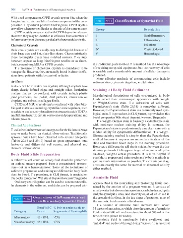
longitudinal axis is parallel to the slow component o the com- TABLE 29.17 Classi cation of Synovial Fluid
pensator. T ey exhibit positive bire ringence. CPPD crystals
are yellow when perpendicular to the axis o the compensator. Group Description
CPPD crystals are associated with CPPD deposition disease.
However, they may be identi ed in e usions rom a number o I Nonin ammatory
inf ammatory joint diseases, particularly rheumatoid arthritis. II In ammatory
Cholesterol Crystals III Infectious
Cholesterol crystals are usually easy to distinguish because o IV Crystal induced
their large size and f at, plate-like shape. Characteristically, V Hemorrhagic
these rectangular plates have notched corners. T ey may,
however, appear as long, bire ringent needles or as rhom-
boids, resembling MSU or CPPD crystals. the traditional push method. T is method has the advantage
T e presence o cholesterol crystals is considered to be o requiring no special equipment, but the recovery o cells
nonspeci c. However, they are usually ound in chronic e u- is variable and a considerable amount o cellular damage is
sions rom patients with rheumatoid arthritis. produced.
More e ective methods o concentrating cells include
Arti acts sedimentation, cytocentri ugation, and ltration.
Artifacts can be mistaken or crystals, although crystals have
sharp, clearly de ned edges and straight sides. Particulate Staining of Body Fluid Sediment
matters that can be con used with crystals include plastic
joint prostheses, nail polish, dust particles, immersion oil Morphological descriptions o cells encountered in body
droplets, and re ractile collagen brils. f uids ref ect their microscopic appearance with Wright
CPPD and MSU crystals can be con used with other bire- or Wright-Giemsa stain. T e coloration o cells with
ringent materials including crystalline anticoagulants, such Papanicolaou’s stain ( able 29.18) is somewhat di erent.
as calcium oxalate, ethylenediaminetetraacetic acid (ED A), However, the Papanicolaou’s stain is a commonly used cyto-
and lithium heparin; certain corticosteroid preparations; and logical stain. Tis procedure, in CLSI ormat, is provided on this
talcum powder. book’s companion Web site at thepoint.lww.com/ urgeon6e.
Te Wright-Giemsa stain is basically a cytoplasmic stain
Clinical Applications with moderate nuclear staining ability. In contrast, the
Papanicolaou’s stain is predominantly a nuclear stain with a
T e distinction between various types o arthritis is not always
easy to make based on clinical observations. raditionally, modest ability or cytoplasmic di erentiation. T e Wright-
synovial f uids have been classi ed into several categories Giemsa staining method is simpler than the Papanicolaou
( ables 29.16 and 29.17) based on gross appearance, total method because it requires no immediate xation o the
leukocyte and di erential cell counts, and physical and slide and there ore ewer steps in the staining procedure.
chemical examinations. However, a di erence in cell size is evident between the two
staining protocols. Cells appear larger when prepared by the
Body Fluid Slide Preparation air-dried, Wright-Giemsa procedure. It is most help ul, i
possible, to prepare and stain specimens by both methods to
A di erential cell count on a body f uid should be per ormed gain as much in ormation as possible. T e criteria or diag-
on stained smears prepared rom a concentrated prepara- nosis are exactly the same or normal or abnormal cells by
tion—not in a hemacytometer. Some o the techniques o either method.
sediment preparation and staining are di erent or body f uids
than or blood. T e procedure, in CLSI ormat, is provided on
this book’s companion Web site at thepoint.lww.com/ urgeon6e. Amniotic Fluid
Ordinary centri ugation can be used to concentrate cellu- Amniotic uid is the nourishing and protecting liquid con-
lar elements in the sediment, and slides can be prepared with tained by the amnion o a pregnant woman. It consists o
mostly water but also contains proteins, carbohydrates, lipids
and phospholipids, urea, and electrolytes, all o which aid in
Comparison of In ammatory and the growth o the etus. In the late stages o gestation, most o
TABLE 29.16
Nonin ammatory Synovial Fluids the amniotic f uid consists o etal urine.
T e volume o amniotic f uid increases until about
Total WBC % Polymorphonuclear 34 weeks o gestation, at which time the amount o amniotic
Category Count Segmented Neutrophils f uid is about 800 mL and is reduced to about 600 mL at the
time o birth (about 40 weeks).
In ammatory >2 × 10 /L >75%
9
Amniotic f uid is continually being swallowed and
9
Nonin ammatory <2 × 10 /L <75% “inhaled” and replaced through being “exhaled.” It is essential