Page 700 - Clinical Hematology_ Theory _ Procedures ( PDFDrive )
P. 700

684            PART 8  ■  Fundamentals of Hematological Analysis


                       SPECIAL HEMATOLOGY PROCEDURES (continued)


                                                                 Early "ring" forms                 Trophozoite                     Schizont                   Gametocytes


                                                            m                                        Not usually                    Not usually
                                                            u
                                                            r                                        seen in                        seen in
                                                            a
                                                            p
                                                            i                                        peripheral                     peripheral
                                                            c
                                                            l                                        blood                          blood
                                                            a
                                                            f
                                                            P.


                                                            e
                                                            l
                                                            a
                                                            v
                                                            o
                                                            P.

                                                            &

                                                            x
                                                            a
                                                            v
                                                            i
                                                            v
                                                            P.


                                                            e
                                                            a
                                                            i
                                                            r
                                                            a
                                                            l
                                                            a
                                                            m
                                                            P.


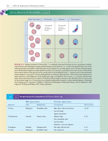
                                 FIGURE 32.9  Malarial parasites in bloo   cells. T is schematic   rawing illustrates the most prominent morpho-

                                 logical  eatures that   istinguish human malarial species in bloo   smears. T e nuclear chromati   bo  ies o  all o  the

                                 malarial parasites are sha  e   in   ark blue-gray in this   iagram but actually appear re   in Giemsa-staine   prepara-
                                 tions. Plasmodium falciparum usually appears as small, f ne, ring  orms, sometimes more than one per re   bloo   cell.

                                 More-mature  orms o  the species are not usually seen in the peripheral bloo  . T e gametocyte is characteristically

                                 banana shape  . P. vivax an   P. ovale are   istinguishe   by   etails not illustrate   here. In ecte   cells an   ring  orms are
                                 larger than those o  P. falciparum. At the trophozoite stage, re   Schu  ner   ots are seen. T e schizont contains more

                                 than a   ozen merozoites be ore it ruptures. P. malariae in ects smaller, senescent cells. Schu  ner   ots are not present.

                                 At the schizont stage, 8 to 12 merozoites are arrange   peripherally aroun   the central malarial pigment. (Reprinte

                                  rom Engleberg NC, Dermo  y   , DiRita V. Schaecter’s Mechanisms of Microbial Disease, 4th e  , Baltimore, MD:

                                 Lippincott Williams & Wilkins, 2007, with permission.)


                    TABLE        32.2        Morphological Comparison of Plasm odium  sp.


                                                           RBC Appearance                                                Parasitic Appearance


                   Species                                 Size                    Inclusions                            Cytoplasm                                                           Merozoites


                   P. vivax                                Enlarged                Schuffner dots                        Blue discs with red nucleus                                         12–24


                                                                                                                         Accole forms

                                                                                                                         Signet ring forms


                   P. falciparum                           Normal                  Maurer dots                           Minute rings                                                        6–32


                                                                                                                         Two chromatic dots


                                                                                                                         Accole forms

                                                                                                                         Gametes: crescent shaped


                   P. malariae                             Normal                  Ziemann dots                          One ring with one dot                                               6–12


                   P. ovale                                Enlarged                Schuffner dots                        One ring form                                                       6–14
   695   696   697   698   699   700   701   702   703   704   705