
Page 438 - Review of Medical Microbiology and Immunology ( PDFDrive )
P. 438
mebooksfree.com
mebooksfree.com
mebooksfree.com
mebooksfree.com
mebooksfree.com mebooksfree.com mebooksfree.com mebooksfree.com mebooksfree.com mebooksfree.com
mebooksfree.com
mebooksfree.com
427
CHAPTER 51 Intestinal & Urogenital—Protozoa
Amebiasis
(Entamoeba histolytica )
2
KEY
I
D
Diagnostic stage
A Infective stage I
Intestinal Disease in Colon
B
mebooksfree.com mebooksfree.com mebooksfree.com mebooksfree.com mebooksfree.com mebooksfree.com
Extraintestinal Disease
B
A
D
1
mebooksfree.com mebooksfree.com mebooksfree.com D mebooksfree.com mebooksfree.com mebooksfree.com
I
D
Cysts and tophozoites
passed in feces
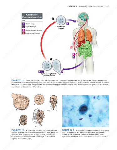
FIGURE 51–1
Entamoeba histolytica. Life cycle. Top blue arrow shows cysts being ingested. Within the intestine, the cyst produces tro-
phozoites that cause amebic dysentery in the colon and can spread to the liver (most often), lung, and brain (Boxes A and B). Bottom blue arrow
shows cysts and trophozoites being passed in the stool and entering the environment. Red arrow indicates survival of cysts in the environment.
(Source: Centers for Disease Control and Prevention.)
mebooksfree.com mebooksfree.com mebooksfree.com mebooksfree.com mebooksfree.com mebooksfree.com
B
A
C
mebooksfree.com mebooksfree.com mebooksfree.com FIGURE 51–3 Entamoeba histolytica—trophozoite. Long arrow mebooksfree.com
mebooksfree.com
mebooksfree.com
D
E
FIGURE 51–2
A: Entamoeba histolytica trophozoite with one
points to trophozoite of E. histolytica. Short arrow points to the
ingested red blood cell and one nucleus (circle with inner dotted line
nucleus of the trophozoite. Arrowhead points to one of the six
represents a red blood cell). B: E. histolytica cyst with four nuclei.
C: Giardia lamblia trophozoite. D: G. lamblia cyst. E: Trichomonas
ingested red blood cells. (Source: Centers for Disease Control and Prevention.)
vaginalis trophozoite (1200×).
mebooksfree.com mebooksfree.com mebooksfree.com mebooksfree.com mebooksfree.com mebooksfree.com