
Page 487 - Review of Medical Microbiology and Immunology ( PDFDrive )
P. 487
mebooksfree.com
mebooksfree.com
mebooksfree.com
mebooksfree.com
mebooksfree.com
mebooksfree.com mebooksfree.com mebooksfree.com Laboratory Diagnosis mebooksfree.com mebooksfree.com
mebooksfree.com
mebooksfree.com
PART VI Parasitology
476
Diagnosis is made microscopically by observing the eggs in
the stools (see Figures 56–3D and 56–12). Occult blood in
the stools is frequent. Eosinophilia is typical.
Treatment
The drug of choice is either albendazole, mebendazole, or
pyrantel pamoate.
mebooksfree.com mebooksfree.com mebooksfree.com Disposing of sewage properly and wearing shoes are effec- mebooksfree.com
mebooksfree.com
mebooksfree.com
Prevention
tive means of prevention.
FIGURE 56–7
Ascaris lumbricoides—egg. Arrow points to an
egg of Ascaris. Note the typical “scalloped” edge of the Ascaris egg.
STRONGYLOIDES
(Source: Public Health Image Library, Centers for Disease Control and Prevention.)
Disease
Strongyloides stercoralis causes strongyloidiasis.
Important Properties
mebooksfree.com mebooksfree.com mebooksfree.com The life cycle of S. stercoralis is shown in Figure 56–13. mebooksfree.com
mebooksfree.com
mebooksfree.com
S. stercoralis has two distinct life cycles, one within the
human body and the other free-living in the soil. The life
cycle in the human body begins with the penetration of
the skin, usually of the feet, by infectious (filariform) lar-
vae (see Figures 56–2I and 56–10) and their migration to
the lungs. They enter the alveoli, pass up the bronchi and
trachea, and then are swallowed. In the small intestine, the
larvae molt into adults (see Figure 56–2H) that enter the
mucosa and produce eggs.
The eggs usually hatch within the mucosa, forming
rhabditiform larvae (see Figure 56–2J) that are passed in
the feces. Some larvae molt to form filariform larvae, which
mebooksfree.com
mebooksfree.com mebooksfree.com mebooksfree.com penetrate the intestinal wall directly without leaving the mebooksfree.com
mebooksfree.com
host and migrate to the lungs (autoinfection). Filariform
larvae can also exit the anus and reinfect through the peri-
anal skin. In immunocompetent patients, this is an infre-
quent, clinically unimportant event.
However, in immunocompromised patients (e.g., those
who have acquired immunodeficiency syndrome [AIDS]
or are taking high-dose corticosteroids or TNF inhibitors)
or patients who are severely malnourished, autoinfection
can lead to massive reinfection (hyperinfection), with
larvae passing to many organs and with severe, sometimes
fatal consequences. Reinfection can also occur in those
infected with human T-cell lymphotropic virus (HTLV)
because their ability to mount a protective T-cell response
mebooksfree.com
mebooksfree.com
mebooksfree.com mebooksfree.com mebooksfree.com and female worms. After mating, the entire life cycle of egg, mebooksfree.com
is diminished.
If larvae are passed in the feces and enter warm, moist
soil, they molt through successive stages to form adult male
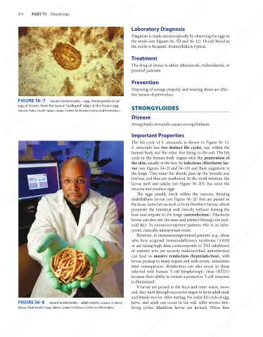
FIGURE 56–8
larva, and adult can occur in the soil. After several free-
Ascaris lumbricoides—adult worms. (Source: Dr. Henry
living cycles, filariform larvae are formed. When they
Bishop, Public Health Image Library, Centers for Disease Control and Prevention.)
mebooksfree.com mebooksfree.com mebooksfree.com mebooksfree.com mebooksfree.com mebooksfree.com