


Page 143 - Textbook of Pathology, 6th Edition
P. 143
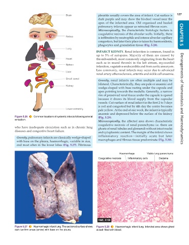
pleuritis usually covers the area of infarct. Cut surface is  127
                                                                 dark purple and may show the blocked vessel near the
                                                                 apex of the infarcted area. Old organised and healed
                                                                 pulmonary infarcts appear as retracted fibrous scars.
                                                                 Microscopically, the characteristic histologic feature is
                                                                 coagulative necrosis of the alveolar walls. Initially, there  CHAPTER 5
                                                                 is infiltration by neutrophils and intense alveolar capillary
                                                                 congestion, but later their place is taken by haemosiderin,
                                                                 phagocytes and granulation tissue (Fig. 5.28).
                                                               INFARCT KIDNEY. Renal infarction is common, found in
                                                               up to 5% of autopsies. Majority of them are caused by
                                                               thromboemboli, most commonly originating from the heart
                                                               such as in mural thrombi in the left atrium, myocardial
                                                               infarction, vegetative endocarditis and from aortic aneurysm.
                                                               Less commonly, renal infarcts may occur due to advanced
                                                               renal artery atherosclerosis, arteritis and sickle cell anaemia.

                                                                 Grossly, renal infarcts are often multiple and may be
                                                                 bilateral. Characteristically, they are pale or anaemic and
                                                                 wedge-shaped with base resting under the capsule and
                                                                 apex pointing towards the medulla. Generally, a narrow
                                                                 rim of preserved renal tissue under the capsule is spared
                                                                 because it draws its blood supply from the capsular  Derangements of Homeostasis and Haemodynamics
                                                                 vessels. Cut surface of renal infarct in the first 2 to 3 days
                                                                 is red and congested but by 4th day the centre becomes
                                                                 pale yellow. At the end of one week, the infarct is typically
                                                                 anaemic and depressed below the surface of the kidney
           Figure 5.26  Common locations of systemic infarcts following arterial  (Fig. 5.29).
           embolism.
                                                                 Microscopically, the affected area shows characteristic
                                                                 coagulative necrosis of renal parenchyma i.e. there are
           who have inadequate circulation such as in chronic lung  ghosts of renal tubules and glomeruli without intact nuclei
           diseases and congestive heart failure.
                                                                 and cytoplasmic content. The margin of the infarct shows
                                                                 inflammatory reaction—initially acute but later
            Grossly, pulmonary infarcts are classically wedge-shaped
            with base on the pleura, haemorrhagic, variable in size,  macrophages and fibrous tissue predominate (Fig. 5.30).
            and most often in the lower lobes (Fig. 5.27). Fibrinous


           Figure 5.27  Haemorrhagic infarct lung. The sectioned surface shows  Figure 5.28  Haemorrhagic infarct lung. Infarcted area shows ghost
           dark tan firm areas (arrow) with base on the pleura.  alveoli filled with blood.

