


Page 147 - Textbook of Pathology, 6th Edition
P. 147
form of injury, the blood flow may be re-established in 3-5  These features, thus, elicit the classical signs of inflam-  131
           seconds while with more severe injury the vasoconstriction  mation—redness, heat, swelling and pain.
           may last for about 5 minutes.
           2. Next follows persistent progressive vasodilatation which  Altered  Vascular Permeability
           involves mainly the arterioles, but to a lesser extent, affects  PATHOGENESIS. In and around the inflamed tissue, there
           other components of the microcirculation like venules and  is accumulation of oedema fluid in the interstitial compart-  CHAPTER 6
           capillaries. This change is obvious within half an hour of  ment which comes from blood plasma by its escape through
           injury. Vasodilatation results in increased blood volume in  the endothelial wall of peripheral vascular bed. In the initial
           microvascular bed of the area, which is responsible for  stage, the escape of fluid is due to vasodilatation and
           redness and warmth at the site of acute inflammation.  consequent elevation in hydrostatic pressure. This is
           3. Progressive vasodilatation, in turn, may elevate the local  transudate in nature. But subsequently, the characteristic
           hydrostatic pressure resulting in transudation of fluid into  inflammatory oedema, exudate, appears by increased
           the extracellular space. This is responsible for swelling at  vascular permeability of microcirculation. The differences
           the local site of acute inflammation.               between transudate and exudate, are already summarised
           4. Slowing or stasis of microcirculation follows which  in Table 4.1 (see page 96).
           causes increased concentration of red cells, and thus, raised  The appearance of inflammatory oedema due to increased
           blood viscosity.                                    vascular permeability of microvascular bed is explained on  Inflammation and Healing
                                                               the basis of Starling’s hypothesis. In normal circumstances,
           5. Stasis or slowing is followed by leucocytic margination
           or peripheral orientation of leucocytes (mainly neutrophils)  the fluid balance is maintained by two opposing sets of forces:
           along the vascular endothelium. The leucocytes stick to the  i) Forces that cause  outward movement of fluid from
           vascular endothelium briefly, and then move and migrate  microcirculation are intravascular hydrostatic pressure and
           through the gaps between the endothelial cells into the  colloid osmotic pressure of interstitial fluid.
           extravascular space. This process is known as emigration  ii) Forces that cause inward movement of interstitial fluid
           (discussed later in detail).                           into circulation are intravascular colloid osmotic pressure and
              The features of haemodynamic changes in inflammation  hydrostatic pressure of interstitial fluid.
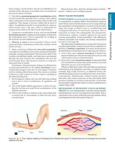
           are best demonstrated by the  Lewis experiment. Lewis  Whatever little fluid is left in the interstitial compartment
           induced the changes in the skin of inner aspect of forearm  is drained away by lymphatics and, thus, no oedema results
           by firm stroking with a blunt point. The reaction so elicited  normally  (Fig. 6.2,A). However, in inflamed tissues, the
           is known as triple response or red line response consisting of  endothelial lining of microvasculature becomes more leaky.
           the following (Fig. 6.1):                           Consequently, intravascular colloid osmotic pressure
           i) Red line appears within a few seconds following stroking  decreases and osmotic pressure of the interstitial fluid
              and is due to local vasodilatation of capillaries and  increases resulting in excessive outward flow of fluid into
              venules.                                         the interstitial compartment which is exudative inflammatory
           ii) Flare is the bright reddish appearance or flush surroun-  oedema (Fig. 6.2,B).
              ding the red line and results from vasodilatation of the  MECHANISMS OF INCREASED VASCULAR PERME-
              adjacent arterioles.                             ABILITY. In acute inflammation, normally non-permeable
           iii) Wheal is the swelling or oedema of the surrounding skin  endothelial layer of microvasculature becomes leaky. This is
              occurring due to transudation of fluid into the extra-  explained by one or more of the following mechanisms which
              vascular space.                                  are diagrammatically illustrated in Fig. 6.3.


           Figure 6.1  A, ‘Triple response’ elicited by firm stroking of skin of forearm with a pencil. B, Diagrammatic view of microscopic features of triple
           response of the skin.

