


Page 144 - Textbook of Pathology, 6th Edition
P. 144
128


     SECTION I


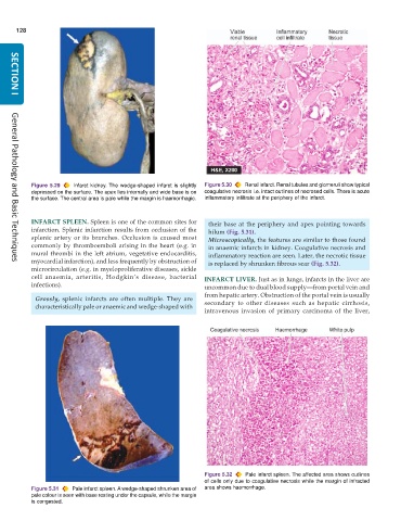
           Figure 5.29  Infarct kidney. The wedge-shaped infarct is slightly  Figure 5.30  Renal infarct. Renal tubules and glomeruli show typical
           depressed on the surface. The apex lies internally and wide base is on  coagulative necrosis i.e. intact outlines of necrosed cells. There is acute
           the surface. The central area is pale while the margin is haemorrhagic.  inflammatory infiltrate at the periphery of the infarct.


           INFARCT SPLEEN. Spleen is one of the common sites for  their base at the periphery and apex pointing towards
           infarction. Splenic infarction results from occlusion of the  hilum (Fig. 5.31).
           splenic artery or its branches. Occlusion is caused most  Microscopically, the features are similar to those found
           commonly by thromboemboli arising in the heart (e.g. in  in anaemic infarcts in kidney. Coagulative necrosis and
           mural thrombi in the left atrium, vegetative endocarditis,  inflammatory reaction are seen. Later, the necrotic tissue
           myocardial infarction), and less frequently by obstruction of  is replaced by shrunken fibrous scar (Fig. 5.32).
     General Pathology and Basic Techniques
           microcirculation (e.g. in myeloproliferative diseases, sickle
           cell anaemia, arteritis, Hodgkin’s disease, bacterial  INFARCT LIVER. Just as in lungs, infarcts in the liver are
           infections).                                        uncommon due to dual blood supply—from portal vein and
                                                               from hepatic artery. Obstruction of the portal vein is usually
            Grossly, splenic infarcts are often multiple. They are
            characteristically pale or anaemic and wedge-shaped with  secondary to other diseases such as hepatic cirrhosis,
                                                               intravenous invasion of primary carcinoma of the liver,


                                                               Figure 5.32  Pale infarct spleen. The affected area shows outlines
                                                               of cells only due to coagulative necrosis while the margin of infracted
           Figure 5.31  Pale infarct spleen. A wedge-shaped shrunken area of  area shows haemorrhage.
           pale colour is seen with base resting under the capsule, while the margin
           is congested.

