


Page 148 - Textbook of Pathology, 6th Edition
P. 148
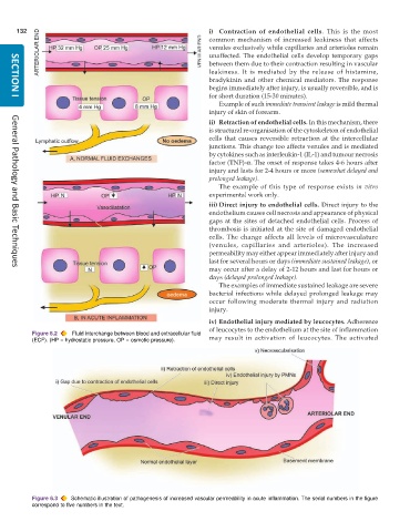
132                                                      i) Contraction of endothelial cells. This is the most
                                                               common mechanism of increased leakiness that affects
                                                               venules exclusively while capillaries and arterioles remain
                                                               unaffected. The endothelial cells develop temporary gaps
                                                               between them due to their contraction resulting in vascular
                                                               leakiness. It is mediated by the release of histamine,
                                                               bradykinin and other chemical mediators. The response
                                                               begins immediately after injury, is usually reversible, and is
                                                               for short duration (15-30 minutes).
     SECTION I
                                                                  Example of such immediate transient leakage is mild thermal
                                                               injury of skin of forearm.
                                                               ii) Retraction of endothelial cells. In this mechanism, there
                                                               is structural re-organisation of the cytoskeleton of endothelial
                                                               cells that causes reversible retraction at the intercellular
                                                               junctions. This change too affects venules and is mediated
                                                               by cytokines such as interleukin-1 (IL-1) and tumour necrosis
                                                               factor (TNF)-α. The onset of response takes 4-6 hours after
                                                               injury and lasts for 2-4 hours or more (somewhat delayed and
                                                               prolonged leakage).
                                                                  The example of this type of response exists  in vitro
                                                               experimental work only.
                                                               iii) Direct injury to endothelial cells. Direct injury to the
                                                               endothelium causes cell necrosis and appearance of physical
                                                               gaps at the sites of detached endothelial cells. Process of
                                                               thrombosis is initiated at the site of damaged endothelial
                                                               cells. The change affects all levels of microvasculature
                                                               (venules, capillaries and arterioles). The increased
                                                               permeability may either appear immediately after injury and
                                                               last for several hours or days (immediate sustained leakage), or
                                                               may occur after a delay of 2-12 hours and last for hours or
     General Pathology and Basic Techniques
                                                               days (delayed prolonged leakage).
                                                                  The examples of immediate sustained leakage are severe
                                                               bacterial infections while delayed prolonged leakage may
                                                               occur following moderate thermal injury and radiation
                                                               injury.
                                                               iv) Endothelial injury mediated by leucocytes. Adherence
                                                               of leucocytes to the endothelium at the site of inflammation
           Figure 6.2  Fluid interchange between blood and extracellular fluid  may result in activation of leucocytes. The activated
           (ECF). (HP = hydrostatic pressure, OP = osmotic pressure).


           Figure 6.3  Schematic illustration of pathogenesis of increased vascular permeability in acute inflammation. The serial numbers in the figure
           correspond to five numbers in the text.

