


Page 19 - Textbook of Pathology, 6th Edition
P. 19
medicine listing 500 remedies, and Sushruta Samhita, similar  Marcello Malpighi (1624–1694) used microscope extensively  3
           book of surgical sciences by Sushruta, and includes about 700  and observed the presence of capillaries and described the
           plant-derived medicines.                            malpighian layer of the skin, and lymphoid tissue in the spleen
              The end of Medieval period was marked by backward  (malpighian corpuscles). Malpighi is known as ‘the father of
           steps in medicine. There were widespread and devastating  histology.’
           epidemics which reversed the process of rational thinking  The credit for beginning of the study of morbid anatomy
           again to supernatural concepts and divine punishment for  (pathologic anatomy), however, goes to Italian anatomist-  CHAPTER 1
           ‘sins.’ The dominant belief during this period was that life  pathologist, Giovanni B. Morgagni (1682–1771). Morgagni was
           was due to influence of vital substance under the control of  an excellent teacher in anatomy, a prolific writer and a
           soul (theory of vitalism). Thus, dissection of human body was  practicing clinician. By his work, Morgagni demolished the
           strictly forbidden as that would mean hurting the ‘soul.’  ancient humoral theory of disease and published his life-time
                                                               experiences based on 700 postmortems and their
           FROM HUMAN ANATOMY TO ERA OF                        corresponding clinical findings. He, thus, laid the foundations
           GROSS PATHOLOGY (AD 1500 to 1800)                   of clinicopathologic methodology in the study of disease and
                                                               introduced the concept of clinicopathologic correlation (CPC),
           The backwardness of Medieval period was followed by the  establishing a coherent sequence of cause, lesions, symptoms,
           Renaissance period i.e. revival of leaning. The Renaissance  and outcome of disease (Fig. 1.3).
           began from Italy in late 15th century and spread to whole of  Sir Percival Pott (1714–1788), famous surgeon in England,  Introduction to Pathology
           Europe. During this period, there was quest for advances in  identified the first ever occupational cancer in the chimney
           art and science. Since there was freedom of thought, there  sweeps in 1775 and discovered chimney soot as the first
           was emphasis on philosophical and rational attitudes again.  carcinogenic agent. However, the study of anatomy in
              The beginning of the development of human anatomy
           took place during this period with the art works and drawings  England during the latter part of 18th Century was
                                                               dominated by the two Hunter brothers: John Hunter (1728–
           of human muscles and embryos by famous Italian painter  1793), a student of Sir Percival Pott, rose to become greatest
           Leonardo da Vinci (1452–1519). Dissection of human body was  surgeon-anatomist of all times and he, together with his elder
           started by Vesalius (1514–1564) on executed criminals. His  brother  William Hunter (1718–1788) who was a reputed
           pupils, Gabriel Fallopius (1523–1562) who described human  anatomist-obstetrician (or man-midwife), started the first
           oviducts (Fallopian tubes) and  Fabricius who discovered  ever museum of pathologic anatomy. John Hunter made a
           lymphoid tissue around the intestine of birds (bursa of  collection of more than 13,000 surgical specimens from his
           Fabricius) further popularised the practice of human anatomic  flourishing practice, arranged them into separate organ
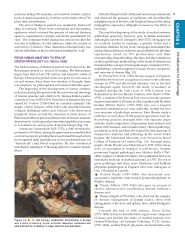
           dissection for which special postmortem amphitheatres came  systems, made comparison of specimens from animals and
           in to existence in various parts of ancient Europe (Fig. 1.2).  plants with humans, and included many clinical pathology
              Antony van Leeuwenhoek (1632–1723), a cloth merchant by
           profession in Holland, during his spare time invented the first  specimens as well, and thus developed the first museum of
                                                               comparative anatomy and pathology in the world which
           ever microscope by grinding the lenses himself through which  became the Hunterian Museum, now housed in Royal
           he recognised male spermatozoa as tiny preformed men (or  College of Surgeons of London (Fig.  1.4). Amongst many
           “homunculi”) and blood corpuscles. He also introduced  pupils of John Hunter was Edward Jenner (1749–1823) whose
           histological staining in 1714 using saffron to examine muscle  work on inoculation in smallpox is well known. Another
           fibres.
                                                               prominent English pathologist was Matthew Baillie (1760–
                                                               1823), nephew of Hunter brothers, who published first-ever
                                                               systematic textbook of morbid anatomy in 1793. The era of
                                                               gross pathology had three more illustrious and brilliant
                                                               physician-pathologists in England who were colleagues at
                                                               Guy’s Hospital in London:
                                                                   Richard Bright (1789–1858) who described non-
                                                               suppurative nephritis, later termed glomerulonephritis or
                                                               Bright’s disease;
                                                                   Thomas Addison (1793–1860) who gave an account of
                                                               chronic adrenocortical insufficiency termed Addison’s
                                                               disease; and
                                                                   Thomas Hodgkin (1798–1866), who observed the complex
                                                               of chronic enlargement of lymph nodes, often with
                                                               enlargement of the liver and spleen, later called Hodgkin’s
                                                               disease.
                                                                  Towards the end of 18th century,  Xavier Bichat
                                                               (1771–1802) in France described that organs were composed
                                                               of tissue and divided the study of morbid anatomy into
           Figure 1.2  In 16th Century, postmortem amphitheatre in Europe  General Pathology and Systemic Pathology. R.T.H. Laennec
           was a place of learning human anatomic dissection conducted and  (1781–1826), another French physician, dominated the early
           demonstrated by professors to eager learners and spectators.

