


Page 21 - Textbook of Pathology, 6th Edition
P. 21
Sir William Leishman (1865–1926) who described Leishman’s  of tumours (Virchow’s lymph node), and components and  5
           stain for blood films in 1914 and observed Leishman-Donovan  diseases of blood (fibrinogen, leukocytosis, leukaemia).
           bodies (LD bodies) in leishmaniasis.                     The concept of frozen section examination when the
              Robert Feulgen (1884–1955) who described Feulgen reaction  patient was still on the operation table was introduced by
           for DNA staining and laid the foundations of cytochemistry and  Virchow’s student,  Julius Cohnheim (1839–1884). In fact,
           histochemistry.                                       during the initial period of development of surgical
              Simultaneous technological advances in machinery   pathology around the turn of the 19th century, frozen  CHAPTER 1
           manufacture led to development and upgradation of     section was considered more acceptable by the surgeons.
           microtomes for obtaining thin sections of organs and tissues  Then there was the period when morphologic examination
           for staining by dyes for enhancing detailed study of sections.  of cells by touch imprint smears was favoured for diagnostic
              Though the presence of cells in thin sections of non-living  purposes than actual tissue sections. Subsequently, further
           object cork had been first demonstrated much earlier by Robert  advances in surgical pathology were made possible by
           Hooke in 1667, it was revived as a unit of living matter in the  improved machinery and development of dyes and stains.
           19th century by  F.T. Schwann (1810–1882), the first     The concept of surgeon and physician doubling up in
           neurohistologist, and Claude Bernarde (1813–1878), pioneer in  the role of pathologist which started in the 19th century
           pathophysiology.                                      continued as late as the middle of the 20th century in most
              Until the end of the 19th century, the study of morbid  clinical departments. Assigning biopsy pathology work to
           anatomy had remained largely autopsy-based and thus had  some faculty member in the clinical department was  Introduction to Pathology
           remained a retrospective science. Rudolf Virchow (1821–1905) in  common practice; that is why some of the notable
           Germany is credited with the beginning of microscopic  pathologists of the first half of 20th century had background
           examination of diseased tissue at cellular level and thus began  of clinical training e.g. James Ewing (1866–1943), A.P. Stout
           histopathology as a method of investigation. Virchow gave two  (1885–1967) and Lauren Ackerman (1905–1993) in US, Pierre
           major hypotheses:                                     Masson (1880–1958) in France, and RA Willis in Australia.
              All cells come from other cells.                      A few other landmarks in further evolution of modern
              Disease is an alteration of normal structure and function of  pathology in this era are as follows:
           these cells.                                              Karl Landsteiner (1863–1943) described the existence of
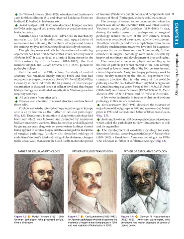
              Virchow came to be referred as Pope in pathology in Europe  major human blood groups in 1900 and was awarded Nobel
           and is aptly known as the ‘father of cellular pathology’  prize in 1930 and is considered father of blood transfusion
           (Fig. 1.6). Thus, sound foundation of diagnostic pathology had  (Fig.  1.7).
           been laid which was followed and promoted by numerous    Ruska and Lorries in 1933 developed electron microscope
           brilliant successive workers. Thus, knowledge and skill gained  which aided the pathologist to view ultrastructure of cell
           by giving accurate diagnosis on postmortem findings started  and its organelles.
           being applied to surgical biopsy and thus emerged the discipline  The development of exfoliative cytology for early
           of surgical pathology. Virchow also described etiology of  detection of cervical cancer began with George N. Papanicolaou
           embolism (Virchow’s triad—slowing of blood-stream, changes  (1883–1962), a Greek-born American pathologist, in 1930s
           in the vessel wall, changes in the blood itself), metastatic spread  who is known as ‘father of exfoliative cytology’ (Fig. 1.8).


             FATHER OF CELLULAR PATHOLOGY        FATHER OF BLOOD TRANSFUSION       FATHER OF EXFOLIATIVE CYTOLOGY


           Figure 1.6  Rudolf Virchow (1821-1905).  Figure 1.7  Carl Landsteiner (1863-1943).  Figure 1.8  George N. Papanicolaou
           German pathologist who proposed cellular  An Austrian pathologist who first discovered the  (1883-1962). American  pathologist,  who
           theory of disease.                 existence of major human blood groups in 1900  developed Pap test for diagnosis of cancer of
                                              and was recipient of Nobel prize in 1930.  uterine cervix.

