


Page 39 - Textbook of Pathology, 6th Edition
P. 39
together measuring about a metre in length in a human                                                    23
           diploid cell. Of these, there are 22 pairs (44 chromosomes) of
           autosomes and one pair of sex chromosomes, either XX (female)
           or XY (male). Each chromosome is composed of two
           chromatids connected at the centromere to form ‘X’
           configuration having variation in location of the centromere.
           Depending upon the length of chromosomes and centromeric                                                   CHAPTER 3
           location, 46 chromosomes are categorised into 7 groups from
           A to G according to Denver classification (adopted at a meeting
           in Denver, USA).
              Chromosomes are composed of 3 components, each with
           distinctive function. These are: deoxyribonucleic acid (DNA)
           comprising about 20%, ribonucleic acid (RNA) about 10%,
           and the remaining 70% consists of nuclear proteins that
           include a number of basic proteins (histones), neutral
           proteins, and acid proteins. DNA of the cell is largely
           contained in the nucleus. The only other place in the cell that
           contains small amount of DNA is mitochondria. Nuclear
           DNA along with histone nuclear proteins form bead-like
           structures called nucleosomes which are studded along the                                                  Cell Injury and Cellular Adaptations
           coils of DNA. Nuclear DNA carries the genetic information
           that is passed via RNA into the cytoplasm for manufacture
           of proteins of similar composition. During cell division, one
           half of DNA molecule acts as a template for the manufacture
           of the other half by the enzyme, DNA polymerase, so that
           the genetic characteristics are transmitted to the next progeny
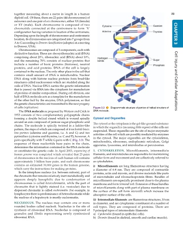
           of cells (replication).                             Figure 3.3  Diagrammatic structure of portion of helical structure of
              The DNA molecule as proposed by Watson and Crick in  DNA molecule.
           1953 consists of two complementary polypeptide chains
           forming a double helical strand which is wound spirally  Cytosol and Organelles
           around an axis composed of pentose sugar-phosphoric acid  The cytosol or the cytoplasm is the gel-like ground substance
           chains. The molecule is spirally twisted in a ladder-like  in which the organelles (meaning little organs) of the cells are
           pattern, the steps of which are composed of 4 nucleotide bases:  suspended. These organelles are the site of major enzymatic
           two purines (adenine and guanine, i.e. A and G) and  two  activities of the cell which are possibly mediated by enzymes
           pyrimidines (cytosine and thymine, i.e. C and T); however, A  in the cytosol. The major organelles are the cytoskeleton,
           pairs specifically with T while G pairs with C (Fig. 3.3). The  mitochondria, ribosomes, endoplasmic reticulum, Golgi
           sequence of these nucleotide base pairs in the chain,  apparatus, lysosomes, and microbodies or peroxisomes.
           determines the information contained in the DNA molecule
           or constitutes the genetic code. In April 2003, sequencing of  1. CYTOSKELETON.  Microfilaments, intermediate
           human genome was completed which revealed that 23 pairs  filaments, and microtubules are responsible for maintaining
           of chromosomes in the nucleus of each human cell contains  cellular form and movement and are collectively referred to
           approximately 3 billion base pairs, and each chromosome  as cytoskeleton.
           contains an estimated 30,000 genes in the human genome,  i) Microfilaments are long filamentous structures having
           which carry the instructions for making proteins.   a diameter of 6-8 nm. They are composed of contractile
              In the interphase nucleus (i.e. between mitosis), part of  proteins, actin and myosin, and diverse materials like parts
           the chromatin that remains relatively inert metabolically and  of microtubules and ribonucleoprotein fibres. Bundles of
           appears deeply basophilic due to condensation of    microfilaments are especially prominent close to the plasma
           chromosomes is called heterochromatin, while the part of  membrane and form terminal web. Extension of these bundles
           chromatin that is lightly stained (i.e. vesicular) due to  of microfilaments along with part of plasma membrane on
           dispersed chromatin is called euchromatin. For example, in  the surface of the cell form microvilli which increase the
           lymphocytes there is predominance of heterochromatin while  absorptive surface of the cells.
           the nucleus of a hepatocyte is mostly euchromatin.
                                                               ii) Intermediate filaments  are filamentous structures, 10 nm
           NUCLEOLUS.  The nucleus may contain one or more     in diameter, and are cytoplasmic constituent of a number of
           rounded bodies called nucleoli. Nucleolus is the site of  cell types. They are composed of proteins. There are
           synthesis of ribosomal RNA. Nucleolus is composed of  5 principal types of intermediate filaments:
           granules and fibrils representing newly synthesised  a) Cytokeratin (found in epithelial cells).
           ribosomal RNA.                                      b) Desmin (found in skeletal, smooth and cardiac muscle).

