


Page 41 - Textbook of Pathology, 6th Edition
P. 41
i) fibrillar structural proteins (collagen, elastin); 25
ii) adhesion proteins (fibronectin, laminin, fibrillin,
osteonectin, tenacin); and
iii) molecules of proteoglycans and glycosaminoglycans
(heparan sulphate, chondroitin sulphate, dermatan sulphate,
keratan sulphate, hyaluronic acid). CHAPTER 3
CAMs participate in fertilisation, embryogenesis, tissue
repair, haemostasis, cell death by apoptosis and in
inflammation. CAMs may be detected on the surface of cells
as well as free in circulation. There are 5 groups of CAMs:
i) Integrins. They have alpha (or CD11*) and beta (CD18)
subunits and have a role in cell-ECM interactions and in
leucocyte-endothelial cell interaction.
ii) Cadherins. These are calcium-dependent adhesion
molecules which bindadjacent cells together and prevent
invasion of ECM by cancer cells. Various types of cadherins
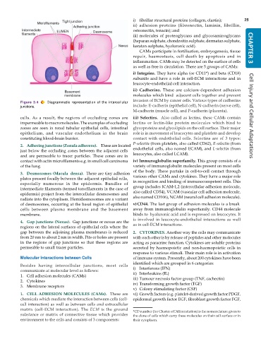
Figure 3.4 Diagrammatic representation of the intercellular
junctions. include: E-cadherin (epithelial cell), N-cadherin (nerve cell),
M-cadherin (muscle cell), and P-cadherin (placenta).
cells. As a result, the regions of occluding zones are iii) Selectins. Also called as lectins, these CAMs contain Cell Injury and Cellular Adaptations
impermeable to macromolecules. The examples of occluding lectins or lectin-like protein molecules which bind to
zones are seen in renal tubular epithelial cells, intestinal glycoproteins and glycolipids on the cell surface. Their major
epithelium, and vascular endothelium in the brain role is in movement of leucocytes and platelets and develop
constituting blood-brain barrier. contact with endothelial cells. Selectins are of 3 types:
P-selectin (from platelets, also called CD62), E-selectin (from
2. Adhering junctions (Zonula adherens). These are located
just below the occluding zones between the adjacent cells endothelial cells, also named ECAM), and L-selectin (from
and are permeable to tracer particles. These zones are in leucocytes, also called LCAM).
contact with actin microfilaments e.g. in small cell carcinoma iv) Immunoglobulin superfamily. This group consists of a
of the lung. variety of immunoglobulin molecules present on most cells
of the body. These partake in cell-to-cell contact through
3. Desmosomes (Macula densa). These are tiny adhesion
plates present focally between the adjacent epithelial cells, various other CAMs and cytokines. They have a major role
especially numerous in the epidermis. Bundles of in recognition and binding of immunocompetent cells. This
intermediate filaments (termed tonofilaments in the case of group includes ICAM-1,2 (intercellular adhesion molecule,
epidermis) project from the intercellular desmosomes and also called CD54), VCAM (vascular cell adhesion molecule,
radiate into the cytoplasm. Hemidesmosomes are a variant also named CD106), NCAM (neural cell adhesion molecule).
of desmosomes, occurring at the basal region of epithelial v)CD44. The last group of adhesion molecules is a break
cells between plasma membrane and the basement away from immunoglobulin superfamily. CD44 molecule
membrane. binds tohyaluronic acid and is expressed on leucocytes. It
is involved in leucocyte-endothelial interactions as well
4. Gap junctions (Nexus). Gap junctions or nexus are the
regions on the lateral surfaces of epithelial cells where the as in cell-ECM interactions.
gap between the adjoining plasma membranes is reduced 2. CYTOKINES. Another way the cells may communicate
from 20 nm to about 2 nm in width. Pits or holes are present with each other is by release of peptides and other molecules
in the regions of gap junctions so that these regions are acting as paracrine function. Cytokines are soluble proteins
permeable to small tracer particles. secreted by haemopoietic and non-haemopoietic cells in
response to various stimuli. Their main role is in activation
Molecular Interactions between Cells of immune system. Presently, about 200 cytokines have been
identified which are grouped in 6 categories:
Besides having intercellular junctions, most cells
communicate at molecular level as follows: i) Interferons (IFN)
1. Cell adhesion molecules (CAMs) ii) Interleukins (IL)
2. Cytokines iii) Tumour necrosis factor group (TNF, cachectin)
iv) Transforming growth factor (TGF)
3. Membrane receptors
v) Colony stimulating factor (CSF)
1. CELL ADHESION MOLECULES (CAMs). These are vi) Growth factors (e.g. platelet-derived growth factor PDGF,
chemicals which mediate the interaction between cells (cell- epidermal growth factor EGF, fibroblast growth factor FGF,
cell interaction) as well as between cells and extracellular
matrix (cell-ECM interaction). The ECM is the ground *CD number (for Cluster of Differentiation) is the nomenclature given to
substance or matrix of connective tissue which provides the clone of cells which carry these molecules on their cell surface or in
environment to the cells and consists of 3 components: their cytoplasm.

