


Page 427 - Textbook of Pathology, 6th Edition
P. 427
begins in the foot and progresses slowly upwards to involve TABLE 15.5: Tumours and Tumour-like Lesions of Blood 411
the whole extremity. With passage of time, the affected area Vessels and Lymphatics.
becomes rough and the oedema is non-pitting. The etiology
is unknown but probably the condition is related to female A. Benign Tumours and Hamartomas
1.
Haemangioma
reproductive system because of preponderance in females 2. Lymphangioma
and aggravation during menses. 3. Glomus tumour (glomangioma)
4. Arteriovenous malformations
II. SECONDARY (OBSTRUCTIVE) LYMPHOEDEMA. 5. Bacillary angiomatosis
This is more common form of lymphoedema. Various causes
of lymphatic obstruction causing lymphoedema are as under: B. Intermediate Grade Tumours
Haemangioendothelioma
i) Lymphatic invasion by malignant tumour.
ii) Surgical removal of lymphatics e.g. in radical C. Malignant Tumours
Haemangiopericytoma
1.
mastectomy. 2. Angiosarcoma
iii) Post-irradiation fibrosis. 3. Kaposi’s sarcoma
iv) Parasitic infestations e.g. in filariasis of lymphatics
producing elephantiasis.
v) Lymphangitis causing scarring and obstruction. A. BENIGN TUMOURS AND HAMARTOMAS
Obstructive lymphoedema occurs only when the Haemangioma
obstruction is widespread as otherwise collaterals develop.
The affected area consists of dilatation of lymphatics distal Haemangiomas are quite common lesions, especially in
to obstruction with increased interstitial fluid. With passage infancy and childhood. The most common site is the skin of
of time, there is inflammatory scarring and the lymphatics the face. Amongst the various clinical and histologic types,
become fibrosed with enlargement of the affected part. three important forms are described below.
Rupture of dilated large lymphatics may result in escape of CAPILLARY HAEMANGIOMA. These are the most
milky chyle into the peritoneum (chyloperitoneum), into the common type. Clinically, they appear as small or large, flat
pleural cavity (chylothorax), into pericardial cavity (chylo- or slightly elevated, red to purple, soft and lobulated lesions, CHAPTER 15
pericardium) and into the urinary tract (chyluria). varying in size from a few millimeters to a few centimeters
in diameter. They may be present at birth or appear in early
TUMOURS AND TUMOUR-LIKE LESIONS childhood. Strawberry birthmarks and ‘port-wine mark’ are
some good examples. The common sites are the skin,
Majority of benign vascular tumours are malformations or subcutaneous tissue and mucous membranes of oral cavity
hamartomas. A hamartoma is a tumour-like lesion made up and lips. Less common sites are internal visceral organs like
of tissues indigenous to the part but lacks the true growth liver, spleen and kidneys.
potential of true neoplasms. However, there is no clear-cut
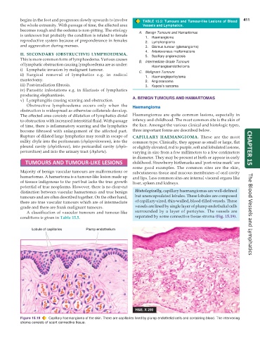
distinction between vascular hamartomas and true benign Histologically, capillary haemangiomas are well-defined
tumours and are often described together. On the other hand, but unencapsulated lobules. These lobules are composed
there are true vascular tumours which are of intermediate of capillary-sized, thin-walled, blood-filled vessels. These
grade and there are frank malignant tumours. vessels are lined by single layer of plump endothelial cells
A classification of vascular tumours and tumour-like surrounded by a layer of pericytes. The vessels are
conditions is given in Table 15.5. separated by some connective tissue stroma (Fig. 15.19). The Blood Vessels and Lymphatics
Figure 15.19 Capillary haemangioma of the skin. There are capillaries lined by plump endothelial cells and containing blood. The intervening
stroma consists of scant connective tissue.

