


Page 430 - Textbook of Pathology, 6th Edition
P. 430
414


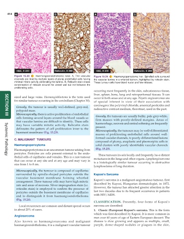
           Figure 15.23  Haemangioendothelioma nose. A, The vascular  Figure 15.24  Haemangiopericytoma liver. Spindled cells surround
           channels are lined by multiple layers of plump endothelial cells having  the vascular lumina in a whorled fashion, highlighted by reticulin stain.
           minimal mitotic activity obliterating the lumina. B, Reticulin stain shows  These tumour cells have bland nuclei and few mitoses.
           condensation of reticulin around the vessel wall but not between the
           proliferating cells.
                                                               occurring most frequently in the skin, subcutaneous tissue,
                                                               liver, spleen, bone, lung and retroperitoneal tissues. It can
           sized and large veins. Haemangioblastoma is the term used  occur in both sexes and at any age. Hepatic angiosarcomas are
           for similar tumour occurring in the cerebellum (Chapter 30).  of special interest in view of their association with
                                                               carcinogens like polyvinyl chloride, arsenical pesticides and
            Grossly, the tumour is usually well-defined, grey-red,  radioactive contrast medium, thorotrast, used in the past.
            polypoid mass.
            Microscopically, there is active proliferation of endothelial  Grossly, the tumours are usually bulky, pale grey-white,
     SECTION III
            cells forming several layers around the blood vessels so  firm masses with poorly-defined margins. Areas of
            that vascular lumina are difficult to identify. These cells  haemorrhage, necrosis and central softening are frequently
            may have variable mitotic activity. Reticulin stain  present.
            delineates the pattern of cell proliferation inner to the  Microscopically, the tumours may be well-differentiated
            basement membrane (Fig. 15.23).
                                                                 masses of proliferating endothelial cells around well-
                                                                 formed vascular channels, to poorly-differentiated lesions
           C. MALIGNANT  TUMOURS
                                                                 composed of plump, anaplastic and pleomorphic cells in
           Haemangiopericytoma                                   solid clusters with poorly identifiable vascular channels
           Haemangiopericytoma is an uncommon tumour arising from  (Fig. 15.25).
           pericytes. Pericytes are cells present external to the endo-  These tumours invade locally and frequently have distant
           thelial cells of capillaries and venules. This is a rare tumour
     Systemic Pathology
           that can occur at any site and at any age and may vary in  metastases in the lungs and other organs. Lymphangiosarcoma
                                                               is a histologically similar tumour occurring in obstructive
           size from 1 to 8 cm.
                                                               lymphoedema of long duration.
            Microscopically, the tumour is composed of capillaries
            surrounded by spindle-shaped pericytes outside the  Kaposi’s Sarcoma
            vascular basement membrane forming whorled         Kaposi’s sarcoma is a malignant angiomatous tumour, first
            arrangement. These tumour cells may have high mitotic
            rate and areas of necrosis. Silver impregnation stain (i.e.  described by Kaposi, Hungarian dermatologist, in 1872.
            reticulin stain) is employed to confirm the presence of  However, the tumour has attracted greater attention in the
            pericytes outside the basement membrane of capillaries  last two decades due to its frequent occurrence in patients
            and to distinguish it from haemangioendothelioma   with HIV/AIDS.
            (Fig. 15.24).
                                                               CLASSIFICATION.  Presently, four forms of Kaposi’s
              Local recurrences are common and distant spread occurs  sarcoma are described:
           in about 20% of cases.                              1. Classic (European) Kaposi’s sarcoma. This is the form
                                                               which was first described by Kaposi. It is more common in
           Angiosarcoma
                                                               men over 60 years of age of Eastern European descent. The
           Also known as haemangiosarcoma and malignant        disease is slow growing and appears as multiple, small,
           haemangioendothelioma, it is a malignant vascular tumour  purple, dome-shaped nodules or plaques in the skin,

