


Page 431 - Textbook of Pathology, 6th Edition
P. 431
415


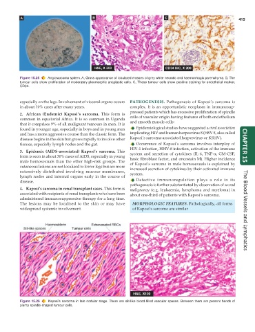
           Figure 15.25  Angiosarcoma spleen. A, Gross appearance of lobulated masses of grey white necrotic and haemorrhagic parenchyma. B, The
           tumour cells show proliferation of moderately pleomorphic anaplastic cells. C, These tumour cells show positive staining for endothelial marker,
           CD34.


           especially on the legs. Involvement of visceral organs occurs  PATHOGENESIS.  Pathogenesis of Kaposi’s sarcoma is
           in about 10% cases after many years.                complex. It is an opportunistic neoplasm in immunosup-
                                                               pressed patients which has excessive proliferation of spindle
           2. African (Endemic) Kaposi’s sarcoma. This form is
           common in equatorial Africa. It is so common in Uganda  cells of vascular origin having features of both endothelium
           that it comprises 9% of all malignant tumours in men. It is  and smooth muscle cells:
           found in younger age, especially in boys and in young men  Epidemiological studies have suggested a viral association
           and has a more aggressive course than the classic form. The  implicating HIV and human herpesvirus 8 (HSV 8, also called
           disease begins in the skin but grows rapidly to involve other  Kaposi’s sarcoma-associated herpesvirus or KSHV).
           tissues, especially lymph nodes and the gut.           Occurrence of Kaposi’s sarcoma involves interplay of
                                                               HIV-1 infection, HHV-8 infection, activation of the immune  CHAPTER 15
           3. Epidemic (AIDS-associated) Kaposi’s sarcoma. This  system and secretion of cytokines (IL-6, TNF-α, GM-CSF,
           form is seen in about 30% cases of AIDS, especially in young  basic fibroblast factor, and oncostain M). Higher incidence
           male homosexuals than the other high-risk groups. The  of Kaposi’s sarcoma in male homosexuals is explained by
           cutaneous lesions are not localised to lower legs but are more  increased secretion of cytokines by their activated immune
           extensively distributed involving mucous membranes,  system.
           lymph nodes and internal organs early in the course of
           disease.                                               Defective immunoregulation plays a role in its
                                                               pathogenesis is further substantiated by observation of second
           4. Kaposi’s sarcoma in renal transplant cases. This form is  malignancy (e.g. leukaemia, lymphoma and myeloma) in
           associated with recipients of renal transplants who have been  about one-third of patients with Kaposi’s sarcoma.
           administered immunosuppressive therapy for a long time.
           The lesions may be localised to the skin or may have  MORPHOLOGIC FEATURES. Pathologically, all forms
           widespread systemic involvement.                      of Kaposi’s sarcoma are similar                      The Blood Vessels and Lymphatics


           Figure 15.26  Kaposi’s sarcoma in late nodular stage. There are slit-like blood-filled vascular spaces. Between them are present bands of
           plump spindle-shaped tumour cells.

