


Page 428 - Textbook of Pathology, 6th Edition
P. 428
412


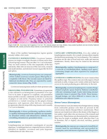
           Figure 15.20  Cavernous haemangioma of the liver. The vascular spaces are large, dilated, many containing blood, and are lined by flattened
           endothelial cells. Scanty connective tissue stroma is seen between the cavernous spaces.


              Many of the capillary haemangiomas regress sponta-  CAPILLARY LYMPHANGIOMA.  It is also called as
           neously within a few years.                         lymphangioma simplex. It is a small, circumscribed, slightly
                                                               elevated lesion measuring 1 to 2 cm in diameter. The common
           CAVERNOUS HAEMANGIOMA.  Cavernous haeman-           locations are the skin of head and neck, axilla and mucous
           giomas are single or multiple, discrete or diffuse, red to blue,  membranes. Rarely, these may be found in the internal
           soft and spongy masses. They are often 1 to 2 cm in diameter.  organs.
           They are most common in the skin (especially of the face
           and neck); other sites are mucosa of the oral cavity, stomach  Histologically, capillary lymphangioma is composed of
     SECTION III
           and small intestine, and internal visceral organs like the liver  a network of endothelium-lined, capillary-sized spaces
           and spleen.                                           containing lymph and often separated by lymphoid
                                                                 aggregates.
            Histologically, cavernous haemangiomas are composed
            of thin-walled cavernous vascular spaces, filled partly or  CAVERNOUS LYMPHANGIOMA. It is more common than
            completely with blood. The vascular spaces are lined by  the capillary type. The common sites are in the region of head
            flattened endothelial cells. They are separated by scanty
            connective tissue stroma (Fig. 15.20).             and neck or axilla. A large cystic variety called cystic hygroma
                                                               occurs in the neck producing gross deformity in the neck.
              Cavernous haemangiomas rarely involute spontaneously.
                                                                 Histologically, cavernous lymphangioma consists of large
           GRANULOMA PYOGENICUM. Granuloma pyogenicum            dilated lymphatic spaces lined by flattened endothelial
     Systemic Pathology
           is also referred to as haemangioma of granulation tissue type.  cells and containing lymph. Scanty intervening stromal
           True to its name, it appears as exophytic, red granulation  connective tissue is present (Fig. 15.21). These lesions,
           tissue just like a nodule, commonly on the skin and mucosa  though benign, are often difficult to remove due to
           of gingiva or oral cavity.  Pregnancy tumour  or granuloma  infiltration into adjacent tissues.
           gravidarum is a variant occurring on the gingiva during
           pregnancy and regresses after delivery. Granuloma   Glomus Tumour (Glomangioma)
           pyogenicum often develops following trauma and is usually  Glomus tumour is an uncommon true benign tumour arising
           1 to 2 cm in diameter.                              from contractile glomus cells that are present in the
                                                               arteriovenous shunts (Sucquet-Hoyer anastomosis). These
            Histologically, it shows proliferating capillaries similar  tumours are found most often in the dermis of the fingers or
            to capillary haemangioma but the capillaries are separated  toes under a nail; other sites are mucosa of the stomach and
            by abundant oedema and inflammatory infiltrate, thus  nasal cavity. These lesions are characterised by extreme pain.
            resembling inflammatory granulation tissue.        They may be single or multiple, small, often less than 1 cm
                                                               in diameter, flat or slightly elevated, red-blue, painful
           Lymphangioma                                        nodules.
           Lymphangiomas are lymphatic counterparts of vascular  Histologically, the tumours are composed of small blood
           angiomas. Lymphangiomas are congenital lesions which are  vessels lined by endothelium and surrounded by
           classified as capillary, cavernous and cystic hygroma.  aggregates, nests and masses of glomus cells. The glomus
           Combinations are also often seen.

