


Page 429 - Textbook of Pathology, 6th Edition
P. 429
413


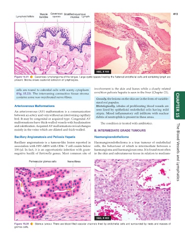
           Figure 15.21  Cavernous lymphangioma of the tongue. Large cystic spaces lined by the flattened endothelial cells and containing lymph are
           present. Stroma shows scattered collection of lymphocytes.


            cells are round to cuboidal cells with scanty cytoplasm  involvement is the skin and bones while a closely related
            (Fig. 15.22). The intervening connective tissue stroma  condition peliosis hepatis is seen in the liver (Chapter 21).
            contains some non-myelinated nerve fibres.
                                                                 Grossly, the lesions on the skin are in the form of variable-
                                                                 sized red papules.
           Arteriovenous Malformations                           Histologically, lobules of proliferating blood vessels are  CHAPTER 15
                                                                 seen lined by epithelioid endothelial cells having mild
           An arteriovenous (AV) malformation is a communication  atypia. Mixed inflammatory cell infiltrate with nuclear
           between an artery and vein without an intervening capillary  debris of neutrophils is present in these areas.
           bed. It may be congenital or acquired type. Congenital AV
           malformations have thick-walled vessels with hyalinisation  The condition is treated with antibiotics.
           and calcification. Acquired AV malformations reveal changes
           mainly in the veins which are dilated and thick-walled.  B. INTERMEDIATE GRADE TUMOURS

           Bacillary Angiomatosis and Peliosis Hepatis         Haemangioendothelioma
           Bacillary angiomatosis is a tumour-like lesion reported in  Haemangioendothelioma is a true tumour of endothelial
           association with HIV-AIDS with CD4+ T cell counts below  cells, the behaviour of which is intermediate between a
           100/μl. In fact, it is an opportunistic infection with gram-  haemangioma and haemangiosarcoma. It is found most often
           negative bacilli of Bartonella genus. Most common site of  in the skin and subcutaneous tissue in relation to medium-  The Blood Vessels and Lymphatics


           Figure 15.22  Glomus tumour. There are blood-filled vascular channels lined by endothelial cells and surrounded by nests and masses of
           glomus cells.

