
Page 811 - Textbook of Pathology, 6th Edition
P. 811
to an incomplete diaphragma sella creating an empty sella. pharyngioma and granular cell tumour (choristoma) are the 795
Other less common causes are Sheehan’s syndrome, other benign pituitary tumours found occasionally.
infarction and scarring in an adenoma, irradiation damage, All pituitary tumours, whether benign or malignant,
or surgical removal of the gland. cause symptoms by following 2 ways:
PITUITARY DWARFISM. Severe deficiency of GH in 1. Pressure effects. These are caused by expansion of the
children before growth is completed results in retarded lesion resulting in destruction of the surrounding glandular
growth and pituitary dwarfism. Most commonly, isolated tissue by pressure atrophy. This causes erosion and
GH deficiency is the result of an inherited autosomal enlargement of sella turcica, upward extension of the tumour
recessive disorder. Less often it may be due to a pituitary damaging the optic chiasma, optic nerves, neurohypophysis
adenoma or craniopharyngioma, infarction and trauma to and adjacent cranial nerves, and rarely, downward extension
the pituitary. The clinical features of inherited cases of into the nasopharynx.
pituitary dwarfism appear after one year of age. These 2. Hormonal effects. Depending upon their cell types,
include proportionate retardation in growth of bones, normal pituitary adenomas produce excess of pituitary hormones
mental state for age, poorly-developed genitalia, delayed and the corresponding clinical syndromes of hyper-
puberty and episodes of hypoglycaemia. Pituitary dwarf pituitarism. Infarction and destruction of adenoma may cause
must be distinguished from hypothyroid dwarf (cretinism) symptoms of hypopituitarism.
in which there is achondroplasia and mental retardation
(page 803).
Pituitary Adenomas
B. Hypofunction of Posterior Pituitary and Adenomas are the most common pituitary tumours. They
Hypothalamus are conventionally classified according to their H & E staining
characteristics of granules into acidophil, basophil and
Insufficiency of the posterior pituitary and hypothalamus is chromophobe adenomas. However, this morphologic
uncommon. The only significant clinical syndrome due to classification is considered quite inadequate because of the
hypofunction of the neurohypophysis and hypothalamus is significant functional characteristics of each type of adenoma
diabetes insipidus. including the chromophobe adenoma, which on H & E CHAPTER 27
DIABETES INSIPIDUS. Deficient secretion of ADH causes staining does not show visible granules. As a result of
diabetes insipidus. The causes of ADH deficiency are: advances in the ultrastructural and immunocytochemical
inflammatory and neoplastic lesions of the hypothalamo- studies, a functional classification of pituitary adenoma has
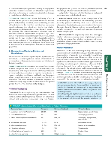
hypophyseal axis, destruction of neurohypophysis due to emerged. Table 27.1 presents a classification of pituitary
surgery, radiation, head injury, and lastly, are those cases adenomas based on functional features as correlated with
where no definite cause is known and are labelled as morphologic features of older classification. The syndromes
idiopathic. The main features of diabetes insipidus are produced by the tumours have been described already.
excretion of a very large volume of dilute urine of low specific
gravity (below 1.010), polyuria and polydipsia. MORPHOLOGIC FEATURES. Grossly, pituitary
adenomas range in size from small foci of less than 10 The Endocrine System
mm in size (termed microadenoma) to large adenomas
PITUITARY TUMOURS several centimeters in diameter. They are spherical, soft
Tumours of the anterior pituitary are more common than and encapsulated.
those of the posterior pituitary and hypothalamus. The most Histologically, by light microscopy of H & E stained
common of the anterior pituitary tumours are adenomas; sections, an adenoma is composed predominantly of one
primary and metastatic carcinomas being rare. Cranio- of the normal cell types of the anterior pituitary i.e.
TABLE 27.1: Morphologic and Functional Classification of Pituitary Adenomas.
Functional Type Frequency Hormones Produced Clinical Syndrome
1. Lactotroph adenoma (Prolactinoma) 20-30% PRL Hypogonadism, galactorrhoea
2. Somatotroph adenoma 5% GH Acromegaly/gigantism
3. Mixed somatotroph-lactotroph adenoma 5% PRL, GH Acromegaly, hypogonadism,
galactorrhoea
4. Corticotroph adenoma 10-15% ACTH Cushing’s syndrome
5. Gonadotroph adenoma 10-15% FSH-LH Inactive or hypogonadism
6. Thyrotroph adenoma 1% TSH Thyrotoxicosis
7. Null cell adenoma/ oncocytoma 20% Nil Pituitary failure
8. Pleurihormonal adenoma 15% Multiple hormones Mixed