
Page 812 - Textbook of Pathology, 6th Edition
P. 812
796 Histologically, craniopharyngioma closely resembles
ameloblastoma of the jaw (page 530). There are 2 distinct
histologic features:
1. Stratified squamous epithelium frequently lining, a
cyst and containing loose stellate cells in the centre; and
2. Solid ameloblastous areas.
Granular Cell Tumour (Choristoma)
Though tumours of the posterior pituitary are rare, granular
cell tumour or choristoma is the most common tumour of
the neurohypophysis. It is composed of a mass of cells having
granular eosinophilic cytoplasm similar to the cells of the
posterior pituitary. It arises as a result of developmental
anomaly and hence the name choristoma. Generally, it
remains asymptomatic and is discovered as an incidental
autopsy finding.
ADRENAL GLAND
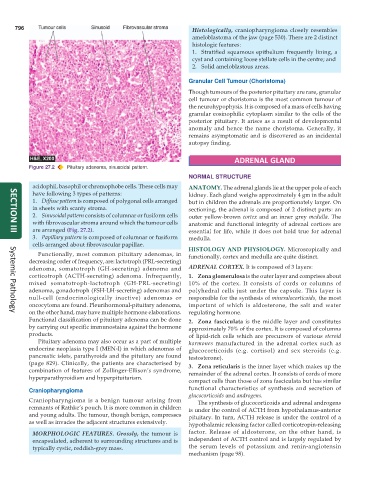
Figure 27.2 Pituitary adenoma, sinusoidal pattern.
NORMAL STRUCTURE
acidophil, basophil or chromophobe cells. These cells may ANATOMY. The adrenal glands lie at the upper pole of each
have following 3 types of patterns: kidney. Each gland weighs approximately 4 gm in the adult
1. Diffuse pattern is composed of polygonal cells arranged but in children the adrenals are proportionately larger. On
in sheets with scanty stroma. sectioning, the adrenal is composed of 2 distinct parts: an
2. Sinusoidal pattern consists of columnar or fusiform cells outer yellow-brown cortex and an inner grey medulla. The
with fibrovascular stroma around which the tumour cells anatomic and functional integrity of adrenal cortices are
are arranged (Fig. 27.2). essential for life, while it does not hold true for adrenal
SECTION III
3. Papillary pattern is composed of columnar or fusiform medulla.
cells arranged about fibrovascular papillae.
HISTOLOGY AND PHYSIOLOGY. Microscopically and
Functionally, most common pituitary adenomas, in functionally, cortex and medulla are quite distinct.
decreasing order of frequency, are: lactotroph (PRL-secreting)
adenoma, somatotroph (GH-secreting) adenoma and ADRENAL CORTEX. It is composed of 3 layers:
corticotroph (ACTH-secreting) adenoma. Infrequently, 1. Zona glomerulosa is the outer layer and comprises about
mixed somatotroph-lactotroph (GH-PRL-secreting) 10% of the cortex. It consists of cords or columns of
adenoma, gonadotroph (FSH-LH-secreting) adenomas and polyhedral cells just under the capsule. This layer is
null-cell (endocrinologically inactive) adenomas or responsible for the synthesis of mineralocorticoids, the most
oncocytoma are found. Pleurihormonal-pituitary adenoma, important of which is aldosterone, the salt and water
on the other hand, may have multiple hormone elaborations. regulating hormone.
Systemic Pathology
Functional classification of pituitary adenoma can be done 2. Zona fasciculata is the middle layer and constitutes
by carrying out specific immunostains against the hormone approximately 70% of the cortex. It is composed of columns
products. of lipid-rich cells which are precursors of various steroid
Pituitary adenoma may also occur as a part of multiple hormones manufactured in the adrenal cortex such as
endocrine neoplasia type I (MEN-I) in which adenomas of glucocorticoids (e.g. cortisol) and sex steroids (e.g.
pancreatic islets, parathyroids and the pituitary are found testosterone).
(page 829). Clinically, the patients are characterised by 3. Zona reticularis is the inner layer which makes up the
combination of features of Zollinger-Ellison’s syndrome, remainder of the adrenal cortex. It consists of cords of more
hyperparathyroidism and hyperpituitarism.
compact cells than those of zona fasciculata but has similar
Craniopharyngioma functional characteristics of synthesis and secretion of
glucocorticoids and androgens.
Craniopharyngioma is a benign tumour arising from The synthesis of glucocorticoids and adrenal androgens
remnants of Rathke’s pouch. It is more common in children is under the control of ACTH from hypothalamus-anterior
and young adults. The tumour, though benign, compresses pituitary. In turn, ACTH release is under the control of a
as well as invades the adjacent structures extensively. hypothalamic releasing factor called corticotropin-releasing
MORPHOLOGIC FEATURES. Grossly, the tumour is factor. Release of aldosterone, on the other hand, is
encapsulated, adherent to surrounding structures and is independent of ACTH control and is largely regulated by
typically cystic, reddish-grey mass. the serum levels of potassium and renin-angiotensin
mechanism (page 98).