
Page 816 - Textbook of Pathology, 6th Edition
P. 816
800 neuromas and von Recklinghausen’s neurofibromatosis in About 10% of pheochromcytomas may be malignant
varying combinations. having tendency for osseous metastases.
The clinical features of pheochromocytoma are
predominantly due to secretion of catecholamines, both Myelolipoma
epinephrine and norepinephrine. The most common feature
is hypertension. Other manifestations due to sudden release Myelolipoma is an uncommon benign adrenal medullary
of catecholamines are congestive heart failure, myocardial tumour found incidentally at autopsy. Less often, it may
infarction, pulmonary oedema, cerebral haemorrhage, and produce symptoms due to excessive hormone elaboration.
even death. The diagnosis is established by measuring 24- MORPHOLOGIC FEATURES. Grossly, a myelolipoma
hour urinary catecholamines or their metabolites such as is usually a small tumour, measuring 0.2-2 cm in diameter.
metanephrine and VMA. Microscopically, it consists of well-differentiated adipose
tissue in which is scattered clumps of haematopoietic cells
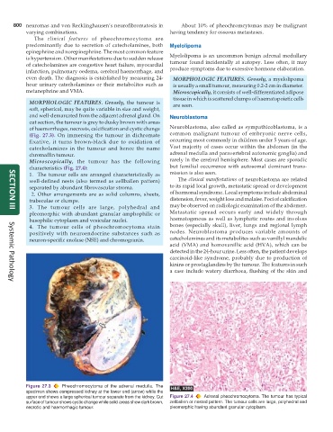
MORPHOLOGIC FEATURES. Grossly, the tumour is are seen.
soft, spherical, may be quite variable in size and weight,
and well-demarcated from the adjacent adrenal gland. On Neuroblastoma
cut section, the tumour is grey to dusky brown with areas
of haemorrhages, necrosis, calcification and cystic change Neuroblastoma, also called as sympathicoblastoma, is a
(Fig. 27.3). On immersing the tumour in dichromate common malignant tumour of embryonic nerve cells,
fixative, it turns brown-black due to oxidation of occurring most commonly in children under 5 years of age.
catecholamines in the tumour and hence the name Vast majority of cases occur within the abdomen (in the
chromaffin tumour. adrenal medulla and paravertebral autonomic ganglia) and
Microscopically, the tumour has the following rarely in the cerebral hemisphere. Most cases are sporadic
characteristics (Fig. 27.4): but familial occurrence with autosomal dominant trans-
1. The tumour cells are arranged characteristically as mission is also seen.
well-defined nests (also termed as zellballen pattern) The clinical manifestations of neuroblastoma are related
separated by abundant fibrovascular stroma. to its rapid local growth, metastatic spread or development
2. Other arrangements are as solid columns, sheets, of hormonal syndrome. Local symptoms include abdominal
trabeculae or clumps. distension, fever, weight loss and malaise. Foci of calcification
3. The tumour cells are large, polyhedral and may be observed on radiologic examination of the abdomen.
SECTION III
pleomorphic with abundant granular amphophilic or Metastatic spread occurs early and widely through
basophilic cytoplasm and vesicular nuclei. haematogenous as well as lymphatic routes and involves
4. The tumour cells of pheochromocytoma stain bones (especially skull), liver, lungs and regional lymph
positively with neuroendocrine substances such as nodes. Neuroblastoma produces variable amounts of
neuron-specific enolase (NSE) and chromogranin. catecholamines and its metabolites such as vanillyl mandelic
acid (VMA) and homovanillic acid (HVA), which can be
detected in the 24-hour urine. Less often, the patient develops
carcinoid-like syndrome, probably due to production of
kinins or prostaglandins by the tumour. The features in such
a case include watery diarrhoea, flushing of the skin and
Systemic Pathology
Figure 27.3 Pheochromocytoma of the adrenal medulla. The
specimen shows compressed kidney at the lower end (arrow) while the
upper end shows a large spherical tumour separate from the kidney. Cut Figure 27.4 Adrenal pheochromocytoma. The tumour has typical
surface of tumour shows cystic change while solid areas show dark brown, zellballen or nested pattern. The tumour cells are large, polyhedral and
necrotic and haemorrhagic tumour. pleomorphic having abundant granular cytoplasm.